File:Red White Blood cells.jpg
此为最大尺寸。
Red_White_Blood_cells.jpg (500 × 326像素,文件大小:57 KB,MIME类型:image/jpeg)
摘要
| 描述 |
العربية: صورة بالميكروسكوب الالكتروني لخلية دم حمراء(يسار), صفيحة دموية(وسط)و خلية دم بيضاء(يمين)
Azərbaycanca: Soldan sağa: eritrosit, trombosit, leykosit
Čeština: Červená krvinka (vlevo), krevní destička (uprostřed) a bílá krvinka (vpravo)
Deutsch: Rasterelektronenmikroskop (REM)-Aufnahme, eingefärbt: Erythrozyt, Thrombozyt, Leukozyt (von links nach rechts)
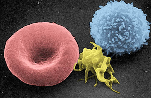
English: Colored Scanning electron microscope (SEM)-picture: erythrocyte, thrombocyte, leukocyte (from left to right:)
Dansk: Fra venstre mod højre: erythrocyt, thrombocyt, leukocyt
Magyar: Színezett Pásztázó elektronmikroszkóp kép: eritrocita, trombocita, leukocita (balról jobbra)
Italiano: Da sinistra verso destra: eritrocita, piastrina e linfocita T
日本語: 左から: 赤血球、血小板、白血球。
Español: De izquierda a derecha: eritrocito, trombocito, linfocito T
Suomi: Väritetty pyyhkäisyelektronimikroskooppikuva, jossa vasemmalta oikealle: punasolu, verihiutale ja valkosolu.
Français : De gauche à droite : érythrocyte, thrombocyte et leucocyte
Nederlands: rode en witte bloedcel, en in het midden een bloedplaatje
Polski: Od lewej do prawej: erytrocyt, trombocyt, leukocyt
Română: De la stânga la dreapta: globulă roşie de sânge, trombocită, limfocită
Русский: Слева направо: эритроцит, тромбоцит, лейкоцит
Português: Da esquerda para a direita : eritrócito, plaqueta e leucócito
中文:由左至右:紅血球,血小板,白血球
A three-dimensional ultrastructural image analysis of a T-lymphocyte (right), a platelet (center) and a red blood cell (left), using a Hitachi S-570 scanning electron microscope (SEM) equipped with a GW Backscatter Detector. |
||||||
| 日期 | 2004年9月21日 (原始上传日期) | ||||||
| 来源 | [1] | ||||||
| 作者 | Electron Microscopy Facility at The National Cancer Institute at Frederick (NCI-Frederick) | ||||||
| 授权 (二次使用本文件) |
|
||||||
| 其他版本 |
此文件衍生的作品: |
||||||

This image was copied from wikipedia:nl where it was uploaded by nl:User:Svdmolen on 21 sep 2004 10:38. The original description was:
rode en witte bloedcel - http://web.ncifcrf.gov/ - PD
文件历史
点击某个日期/时间查看对应时刻的文件。
| 日期/时间 | 缩略图 | 大小 | 用户 | 备注 | |
|---|---|---|---|---|---|
| 当前 | 2016年6月3日 (五) 16:07 | | 500 × 326(57 KB) | Jakob Suckale | Scanning EM picture was colorized to highlight the different cell types. Red blood cell in red, platelet in yellow, lymphocyte in blue. |
| 2016年6月3日 (五) 16:05 |  | 500 × 326(57 KB) | Jakob Suckale | Scanning EM picture was colorized to highlight the different cell types. Red blood cell in red, platelet in yellow, lymphocyte in blue. | |
| 2005年11月9日 (三) 20:44 |  | 500 × 326(36 KB) | E rulez | This image was copied from wikipedia:nl. The original description was: rode en witte bloedcel - http://web.ncifcrf.gov/ - PD {| border="1" ! date/time || username || edit summary |---- | 21 sep 2004 10:38 || Svdmolen || <nowiki>(rode en witte bloedcel - |
文件用途
全域文件用途
以下其他wiki使用此文件:
- als.wikipedia.org上的用途
- ang.wikipedia.org上的用途
- arc.wikipedia.org上的用途
- ar.wikipedia.org上的用途
- ar.wikiversity.org上的用途
- arz.wikipedia.org上的用途
- ast.wikipedia.org上的用途
- be.wikipedia.org上的用途
- bg.wikipedia.org上的用途
- bn.wikipedia.org上的用途
- bn.wikibooks.org上的用途
- bs.wikipedia.org上的用途
- ca.wikipedia.org上的用途
- ckb.wikipedia.org上的用途
- cs.wikipedia.org上的用途
- da.wikipedia.org上的用途
- de.wikipedia.org上的用途
查看本文件的更多全域用途。