File:Schizophrenia PET scan.jpg
此为最大尺寸。
Schizophrenia_PET_scan.jpg (224 × 248像素,文件大小:23 KB,MIME类型:image/jpeg)
摘要
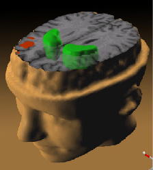
illustration of Schizophrenia's effect on the brain; taken from here archive copy at the Wayback Machine
- Source: Andreas Meyer-Lindenberg, M.D., Ph.D., NIMH Clinical Brain Disorders Branch on the tree of life.
While patients performed a working memory task, the less the prefrontal cortex (red) activated, the more dopamine increased in the striatum (green).
Abstract of study is here.
许可协议
此圖像為美國衛生與公眾服務部下屬國家衛生院(NIH)僱員於公務中所拍攝或製作之作品。作為美國聯邦政府的作品,此圖像屬於公有領域。
 |
||
| 本文件已被确认为免除已知的著作权法限制(包括所有相关权利)。 | ||
文件历史
点击某个日期/时间查看对应时刻的文件。
| 日期/时间 | 缩略图 | 大小 | 用户 | 备注 | |
|---|---|---|---|---|---|
| 当前 | 2005年11月30日 (三) 12:07 | | 224 × 248(23 KB) | Skagedal | illustration of Schizophrenia's effect on the brain; taken [http://www.nih.gov/news/pr/jan2002/nimh-28.htm from here] *Source: Andreas Meyer-Lindenberg, M.D., Ph.D., NIMH Clinical Brain Disorders Branch ''While patients performed a working memory t |
文件用途
以下页面使用本文件:
全域文件用途
以下其他wiki使用此文件:
- ar.wikipedia.org上的用途
- ast.wikipedia.org上的用途
- bg.wikipedia.org上的用途
- ca.wikipedia.org上的用途
- cs.wikipedia.org上的用途
- el.wikipedia.org上的用途
- en.wikipedia.org上的用途
- eo.wikipedia.org上的用途
- es.wikipedia.org上的用途
- fr.wikipedia.org上的用途
- he.wikipedia.org上的用途
- hi.wikipedia.org上的用途
- hy.wikipedia.org上的用途
- id.wikipedia.org上的用途
- kn.wikipedia.org上的用途
- mzn.wikipedia.org上的用途
- no.wikipedia.org上的用途
- pl.wikipedia.org上的用途
- pt.wikipedia.org上的用途
- ru.wikipedia.org上的用途
- sk.wikipedia.org上的用途
- sv.wikipedia.org上的用途
- ta.wikipedia.org上的用途
- tr.wikipedia.org上的用途
查看此文件的更多全域用途。