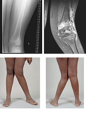
膝外翻
膝外翻(英语:Genu valgum),又名X型腿,是指双腿伸直时膝关节形成一个角度使两膝相互接触。严重的外翻畸形通常使得双腿同时伸直的时候让两侧足部无法相互接触。外翻意味着膝关节的远端(即小腿)向外弯曲,这种情况下膝关节及其近端(即大腿)看起来是向内弯曲的。
| 膝外翻 | |
|---|---|
 | |
| 骨癌治疗后左膝严重的膝外翻 | |
| 类型 | knee disorder[*]、valgus deformity[*] |
| 分类和外部资源 | |
| 医学专科 | 医学遗传学 |
| ICD-10 | M21.0 |
| DiseasesDB | 29408 |
| MedlinePlus | 001263 |
| eMedicine | 1259772 |
站立且两膝彼此接触时如果两侧足部也能彼此接触,属于轻度的膝外翻,常见于2至5岁之间的儿童,通常会随着成长而自然恢复正常。但状况也可能随着年龄增长而持续或恶化,特别是如果有相关疾病,比如佝偻病。先天性或未知原因者称为“特发性膝外翻”。
其他全身性疾病也可能相关,例如施奈德结晶状角膜营养不良,这是一种常伴有高血脂症的染色体显性疾病。
病因
编辑诊断
编辑外翻程度在临床上可透过“Q角”(英语:Q angle)来估算,Q角是由髂骨前上棘到髌骨的线和髌骨到胫骨结节的线所形成的夹角角度。女性膝伸展时Q角应小于22度,膝弯曲90度时Q角应小于9度。男性膝伸展时Q角应小于18度,膝弯曲90度时Q角应小于8度。男性典型Q角为12度,女性为17度[7]。
放射影像检查
编辑利用投射放射影像(X光检查),内翻或外翻畸形的程度可以通过髋膝踝角[8]来量化,该角是股骨机械轴线与踝关节中心之间的夹角角度[9]。成人的正常角度为内翻1.0°到1.5°之间[10]。儿童的正常角度随年龄而异[11]。
-
髋膝踝角
治疗
编辑膝外翻的人常内侧足弓塌陷,使内踝低于外踝。膝外翻的成年人容易受伤并出现慢性膝关节疾病,例如髌骨软骨软化症和骨关节炎,后续可导致严重的疼痛和行走问题。
对于儿童来说,两到五岁之间有膝外翻是正常的,长大后几乎所有外翻都会消失。如果症状持续或来自遗传,医生常会开立夜用矫正鞋或小腿支架逐步将腿移回正位。如果病情持续或恶化,可能需要手术以处理疼痛和并发症[12]。可用的外科手术方法包括下段股骨调整和全膝关节置换术。
减轻体重和使用低冲击运动取代高冲击运动可以帮助减缓病程进展。每走一步,病患体重会对外翻的膝关节施加扭力,其效应随着外翻角度增加或体重增加而增加。
复健科医师或物理治疗师可帮助病患学习如何妥善使用腿部肌肉支撑骨骼结构并改善病情。
较罕见的,某些膝外翻的骨畸形被追踪诊断是由于缺乏骨骼生长所必需的营养素,比如佝偻病(缺乏骨骼营养,尤其是饮食中的维生素D和钙)[1]或坏血病(缺乏维生素C)。治疗其根本的维生素缺乏[12]可帮助回复正常骨骼发育。
参见
编辑参考文献
编辑- ^ 1.0 1.1 NHS. Knock Knees. January 2016 [2019-11-11]. (原始内容存档于2018-01-29).
- ^ Genu Valgum Due to Fluoride Toxicity. Nutrition Reviews. 1975-03-01, 33 (3): 76–77. ISSN 0029-6643. doi:10.1111/j.1753-4887.1975.tb06023.x (英语).
- ^ A Study on Crippling in Skeletal Fluorosis (PDF). [2019-11-11]. (原始内容存档 (PDF)于2021-03-25).
Rigidity of Neck and Restricted Movements of Skull, Kyphosis of thoracic vertebrae, Scoliosis in the chest, bending downwards to see the floor without seeing the sky, criss cross walking, Joint pains in the upper and lower extremities, Genuvarum with bowing of leg, Crippling state of patient without movement, Paraesthesia and Paraplegia are the findings recorded.
- ^ Endemic Fluorosis with Genu Valgum Syndrome in a Village of District Mandla, Madhya Pradesh.
- ^ A clinical and biochemical study of chronic fluoride toxicity in children of Kheru Thanda of Gulbarga district, Karnataka, India.
Radiographic changes suggestive of osteoporosis, osteosclerosis, and genu valgum was observed. Major skeletal manifestations observed in various studies of fluorosis are bowed legs(genu varum) or knock-knee (genu valgum), and stiffness of the cervical andlumbar spine.Our study also revealed skeletal fluorosis with cripplingbone deformities
- ^ Studies in Endemic Genu Valgum — A Manifestation of Fluoride Toxicity in India. [2019-11-11]. (原始内容存档于2018-06-13).
The latter is characterized by the presence of genu valgum, dental fluorosis, osteosclerosis of the spine and simultaneous occurrence of osteoporosis of some other bones such as the lower limb bones.
- ^ Mohammad-Jafar Emami, Mohammad-Hossein Ghahramani, Farzad Abdinejad and Hamid Namazi. Q-angle: an invaluable parameter for evaluation of anterior knee pain. Archives of Iranian medicine. January 2007, 10 (1): 24–26. PMID 17198449.
- ^ W-Dahl, Annette; Toksvig-Larsen, Sören; Roos, Ewa M. Association between knee alignment and knee pain in patients surgically treated for medial knee osteoarthritis by high tibial osteotomy. A one year follow-up study. BMC Musculoskeletal Disorders. 2009, 10 (1): 154. ISSN 1471-2474. PMC 2796991 . PMID 19995425. doi:10.1186/1471-2474-10-154.
- ^ Cherian, Jeffrey J.; Kapadia, Bhaveen H.; Banerjee, Samik; Jauregui, Julio J.; Issa, Kimona; Mont, Michael A. Mechanical, Anatomical, and Kinematic Axis in TKA: Concepts and Practical Applications. Current Reviews in Musculoskeletal Medicine. 2014, 7 (2): 89–95. ISSN 1935-973X. PMC 4092202 . PMID 24671469. doi:10.1007/s12178-014-9218-y.
- ^ Sheehy, L.; Felson, D.; Zhang, Y.; Niu, J.; Lam, Y.-M.; Segal, N.; Lynch, J.; Cooke, T.D.V. Does measurement of the anatomic axis consistently predict hip-knee-ankle angle (HKA) for knee alignment studies in osteoarthritis? Analysis of long limb radiographs from the multicenter osteoarthritis (MOST) study. Osteoarthritis and Cartilage. 2011, 19 (1): 58–64. ISSN 1063-4584. PMC 3038654 . doi:10.1016/j.joca.2010.09.011.
- ^ 11.0 11.1 Sabharwal, Sanjeev; Zhao, Caixia. The Hip-Knee-Ankle Angle in Children: Reference Values Based on a Full-Length Standing Radiograph. The Journal of Bone and Joint Surgery, American Volume. 2009, 91 (10): 2461–2468. ISSN 0021-9355. doi:10.2106/JBJS.I.00015.
- ^ 12.0 12.1 Peter M Stevens. Pediatric Genu Valgum Treatment & Management. eMedicine. 2019-01-03 (英语).
外部链接
编辑- Treating knock knee (页面存档备份,存于互联网档案馆) - UK NHS