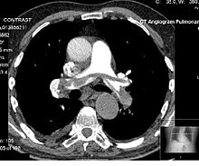
显影计算机断层成像
显影计算机断层成像(英语:contrast CT)指针对受检者施打显影剂的计算机断层成像。计算机断层成像使用的显影剂以含碘显影剂为主,这种方式除有助于区分血管等组织和周边其他组织,也有助于分析组织的功能是否正常。[1]多数情况下,在对受检者施打显影剂前后皆会分别进行成像,显影剂施打前的图像又称为显影前图像(precontrast image)或一般阶段图像(native-phase image),施打后的图像则称为显影后图像(postcontrast image)。[2]


团注追踪
编辑团注追踪(bolus tracking)是一种透过充分把关显影剂的时效以实现显影效果最佳化的方法。带有放射不透明的小型显影剂团注透过周边静脉导管注入受检者体内。依欲成像的血管不同,首先将欲成像的区域周边划为感兴趣区域(region of interest,简称R.O.I.),待显影剂通过该区域时再进行成像,成像的速率应和显影剂于血管中流动的速度保持一致。[3]
显影消退
编辑显影消退(washout)是指显影剂在动脉相阶段被组织吸收后,于静脉相阶段等后续阶段逐渐消退的现象。例如,肝细胞癌的显影计算机断层成像中,透过分析肿瘤部位和肝实质部位显影的时间差,可判断肿瘤的大小和位置。[4]
阶段
编辑依检查目的不同,施打显影剂和成像之间的时间差存在不同的规范,以使不同的器官和组织能达到最佳的显影效果。[5]以下表格列出显影计算机断层成像的主要阶段:[6]
| 阶段 | 显影剂注射后计时[6] | 团注追踪开始后计时[6] | 目标组织和发现[6] |
|---|---|---|---|
| 非显影电脑断层扫描 | - | - | |
| 肺动脉相阶段 | 6-13秒[7] | - | |
| 肺静脉相阶段 | 17-24秒[7] | - | |
| 早期系统性动脉相阶段 | 15-20秒 | 即时 |
|
| 晚期系统性动脉相阶段 又称“动脉相阶段”(arterial phase) 或“早期门静脉相阶段”(early venous portal phase) |
35-40秒 | 15-20秒 |
|
| 胰脏阶段 | 30秒[9]或40-50秒[10] | 20-30秒 | |
| 肝脏阶段(较为准确的说法) 又称“晚期门静脉相阶段”(late venous portal phase) |
70-80秒 | 50-60秒 |
|
| 肾脏阶段 | 100秒 | 80秒 | |
| 系统性静脉相阶段 | 180秒 | 160秒 |
|
| 延迟阶段 又称“显影消退阶段”(wash out phase) 或“均衡阶段”(equilibrium phase) |
6-15分[6] | 6-15分[6] |
|
血管摄影
编辑电脑断层血管摄影为显影计算机断层成像的一种,其针对目标血管的部位和血流相进行成像,以检查血管疾病。例如,腹部主动脉血管摄影(abdominal aortic angiography)是在动脉相阶段(显影剂于动脉中的浓度高峰期)针对腹部的显影,可用于检查如主动脉剥离等症状。[11]
显影剂量
编辑成人
编辑下表显示了正常体重成年人的剂量。 但是,对于有碘显影剂风险的患者,如过敏反应,显影剂诱发的肾病,对甲状腺功能的影响或药物不良相互作用,可能需要调整甚至停止使用剂量。
| 检查项目 | 碘浓度 | 说明 | |||
|---|---|---|---|---|---|
| 300 mg/ml | 350 mg/ml | 370 mg/ml | |||
| 脑部CT | 95 ml[12] | 80 ml[12] | 75 ml[12] | ||
| 肺部CT | 整体 | 70-95 ml[注 1] | 60-80 ml[注 1] | 55-75 ml[注 1] | 要检查出肺部中薄壁组织的变化,往往无需施打静脉注射显影剂 |
| 肺血管CT | 20 ml[注 2] | 17 ml[注 2] | 15 ml[注 2] | 使用特定的低对比分辨率CT时的最低剂量[注 2] | |
| 腹部和骨盆的CT | 整体 | 70 ml[12] | 60 ml[12] | 55 ml[12] | |
| 肝 | 55 ml[注 3] | 45 ml[注 3] | 40-45 ml[注 3] | 最低有效剂量[注 3] | |
| 血管CT | 25 ml[注 4] | 20 ml[注 4] | 使用特定的低对比分辨率CT[注 4] | ||
当受检者的体重不在正常范围内,显影剂的剂量应随之调整,调整幅度应同受检者的除脂肪体重等比。对于较为肥胖的受检者(身体质量指数介于35至40之间),建议使用布尔公式(Boer formula)计算其除脂肪体重:[13]
男性:
女性:
| 缩写 | 中文名称 | 单位 |
|---|---|---|
| LBW | 除脂肪体重 | 公斤 |
| W | 体重 | 公斤 |
| H | 身高 | 米 |
儿童
编辑用于儿童的标准剂量:[14]
| 检查项目 | 碘浓度 | |
|---|---|---|
| 300 mg/ml | 350 mg/ml | |
| 一般检查 | 2.0 ml/kg | 1.7 ml/kg |
| 脑部、颈部或胸部的CT | 1.5 ml/kg | 1.3 ml/kg |
副作用
编辑含碘显影剂除可能引起过敏反应、显影剂肾病变、甲状腺功能亢进症等症状,也可能造成二甲双胍堆积。由于含碘显影剂没有绝对的禁忌症,因此使用前应评估利弊和风险。[15]
就如同其他种类的计算机断层成像一样,辐射剂量增加意味着放射性癌症的风险提高。
注射含碘显影剂有时会造成显影剂外渗。[16]
参见
编辑注释
编辑- ^ 1.0 1.1 1.2 0.3-0.4 gI/kg,用于体重70公斤的成年人,参考来源:
- Iezzi, Roberto; Larici, Anna Rita; Franchi, Paola; Marano, Riccardo; Magarelli, Nicola; Posa, Alessandro; Merlino, Biagio; Manfredi, Riccardo; Colosimo, Cesare. Tailoring protocols for chest CT applications: when and how?. Diagnostic and Interventional Radiology. 2017, 23 (6): 420–427. ISSN 1305-3825. PMC 5669541 . PMID 29097345. doi:10.5152/dir.2017.16615.
- ^ 2.0 2.1 2.2 2.3 使用双能量计算机断层成像(如:90/150 SnkVp),参考来源:
- Leroyer, Christophe; Meier, Andreas; Higashigaito, Kai; Martini, Katharina; Wurnig, Moritz; Seifert, Burkhardt; Keller, Dagmar; Frauenfelder, Thomas; Alkadhi, Hatem. Dual Energy CT Pulmonary Angiography with 6g Iodine—A Propensity Score-Matched Study. PLOS ONE. 2016, 11 (12): e0167214. ISSN 1932-6203. PMC 5132396 . PMID 27907049. doi:10.1371/journal.pone.0167214.
- ^ 3.0 3.1 3.2 3.3 肝脏的CT成像建议使用显影剂,以达到至少30 HU的阻射率,参考来源:
- Multislice CT 3. Springer-Verlag Berlin and Heidelberg GmbH & Co. KG. 2010. ISBN 9783642069680.
- Bae, Kyongtae T. Intravenous Contrast Medium Administration and Scan Timing at CT: Considerations and Approaches. Radiology. 2010, 256 (1): 32–61. ISSN 0033-8419. PMID 20574084. doi:10.1148/radiol.10090908.
- ^ 4.0 4.1 4.2 电脑断层血管摄影,受检者为体重70公斤的成年人,配合CT成像的时间,每公斤体重含碘100-150 mg,管电压为80 kVp(低管电压),固定对比噪声比并使用毫安秒补偿,显影剂施打时间的长度固定,开启自动团注追踪和盐水追踪,参考来源:
- Nyman, Ulf. Contrast Medium-Induced Nephropathy (CIN) Gram-Iodine/GFR Ratio to Predict CIN and Strategies to Reduce Contrast Medium Doses. 2012. doi:10.5772/29992.
参考文献
编辑- ^ Webb, W. Richard; Brant, Wiliam E.; Major, Nancy M. Fundamentals of Body CT. Elsevier Health Sciences. 2014: 152. ISBN 9780323263580 (英语).
- ^ Dahlman, P.; Semenas, E.; Brekkan, E.; Bergman, A.; Magnusson, A. Detection and Characterisation of Renal Lesions by Multiphasic Helical CT. Acta Radiologica. 2000, 41 (4): 361–366. PMID 10937759. doi:10.1080/028418500127345479.
- ^ Kurokawa, Ryo; Maeda, Eriko; Mori, Harushi; Amemiya, Shiori; Sato, Jiro; Ino, Kenji; Torigoe, Rumiko; Abe, Osamu. Effect of bolus tracking region-of-interest position within the descending aorta on luminal enhancement of coronary arteries in coronary computed tomography angiography. Medicine (Baltimore). 2019, 98 (19). PMID 31083207. doi:10.1097/MD.0000000000015538.
- ^ Choi, Jin-Young; Lee, Jeong-Min; Sirlin, Claude B. CT and MR Imaging Diagnosis and Staging of Hepatocellular Carcinoma: Part II. Extracellular Agents, Hepatobiliary Agents, and Ancillary Imaging Features. Radiology. 2014, 273 (1): 30–50. ISSN 0033-8419. PMC 4263770 . PMID 25247563. doi:10.1148/radiol.14132362.
- ^ Bae, Kyongtae T. Intravenous Contrast Medium Administration and Scan Timing at CT: Considerations and Approaches. Radiology. 2010, 256 (1): 32–61. ISSN 0033-8419. PMID 20574084. doi:10.1148/radiol.10090908.
- ^ 6.0 6.1 6.2 6.3 6.4 6.5 Robin Smithuis. CT contrast injection and protocols. Radiology Assistant. [2017-12-13]. (原始内容存档于2019-09-29).
- ^ 7.0 7.1 Page 584 in: Ákos Jobbágy. 5th European Conference of the International Federation for Medical and Biological Engineering 14 - 18 September 2011, Budapest, Hungary. Volume 37 of IFMBE Proceedings. Springer Science & Business Media. 2012. ISBN 9783642235085.
- ^ Pavan Nandra. Introducing the use of Flash CTPA; how does it compare to standard CTPA?. Postering. 2018 [2021-01-08]. (原始内容存档于2020-02-21).
- ^ Raman SP, Fishman EK. Advances in CT Imaging of GI Malignancies.. Gastrointest Cancer Res. 2012, 5 (3 Suppl 1): S4–9. PMC 3413036 . PMID 22876336.
- ^ 10.0 10.1 Otto van Delden and Robin Smithuis. Pancreas - Carcinoma. Radiology Assistant. [2017-12-15]. (原始内容存档于2019-09-26).
- ^ Mirvis, Stuart E.; Soto, Jorge A.; Shanmuganathan, Kathirkamanathan; Yu, Joseph; Kubal, Wayne S. Problem Solving in Emergency Radiology E-Book. Elsevier Health Sciences. 2014: 424. ISBN 9781455758395.
- ^ 12.0 12.1 12.2 12.3 12.4 12.5 New Zealand Datasheet (PDF). New Zealand Medicines and Medical Devices Safety Authority. [2018-10-16]. (原始内容存档 (PDF)于2021-02-22).
- ^ Caruso, Damiano; De Santis, Domenico; Rivosecchi, Flaminia; Zerunian, Marta; Panvini, Nicola; Montesano, Marta; Biondi, Tommaso; Bellini, Davide; Rengo, Marco; Laghi, Andrea. Lean Body Weight-Tailored Iodinated Contrast Injection in Obese Patient: Boer versus James Formula. BioMed Research International. 2018, 2018: 1–6. ISSN 2314-6133. PMC 6110034 . PMID 30186869. doi:10.1155/2018/8521893.
- ^ Nievelstein, Rutger A. J.; van Dam, Ingrid M.; van der Molen, Aart J. Multidetector CT in children: current concepts and dose reduction strategies. Pediatric Radiology. 2010, 40 (8): 1324–1344. ISSN 0301-0449. PMC 2895901 . PMID 20535463. doi:10.1007/s00247-010-1714-7.
- ^ Stacy Goergen. Iodine-containing contrast medium. InsideRadiology - The Royal Australian and New Zealand College of Radiologists. 2017-07-26 [2019-02-22]. (原始内容存档于2021-03-03).
- ^ Hrycyk J, Heverhagen JT, Böhm I. What you should know about prophylaxis and treatment of radiographic and magnetic resonance contrast medium extravasation. Acta Radiol. 2019, 60 (4): 496–500. PMID 29896979. doi:10.1177/0284185118782000.