Gray1144_zh.png(564 × 550像素,文件大小:236 KB,MIME类型:image/png


摘要

描述
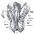
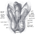
English: The scrotum. The penis has been turned upward, and the anterior wall of the scrotum has been removed. On the right side, the spermatic cord, the infundibuliform fascia, and the Cremaster muscle are displayed; on the left side, the infundibuliform fascia has been divided by a longitudinal incision passing along the front of the cord and the testicle, and a portion of the parietal layer of the tunica vaginalis has been removed to display the testicle and a portion of the head of the epididymis, which are covered by the visceral layer of the tunica vaginalis. (Toldt.)
日期 (UTC)
来源 本文件衍生自: Gray1144.png
作者
其他版本
这是一张修改过的图片,这意味着它已在原版本的基础上通过软件进行了编辑,改动内容:Translated to Chinese。其原始版本为:Gray1144.png。修改者:GnolizX

许可协议

Public domain

本作品作者于PD-scan年逝世,在其来源国以及其他著作权期限是作者逝世后100年或以下的国家和地区属于公有领域


这个作品在美利坚合众国属于公有领域,因为它首次发布早于1929年1月1日,或于该日期之前在美国著作权局登记。

原始上传日志

This image is a derivative work of the following images:

  • File:Gray1144.png licensed with PD-old-100-expired, PD-scan
    • 2007-01-23T19:26:34Z Pngbot 564x550 (78990 Bytes) optimized with optipng
    • 2006-02-11T05:21:24Z Arcadian 564x550 (129950 Bytes) {{Gray's Anatomy plate}}

Uploaded with derivativeFX

说明

添加一行文字以描述该文件所表现的内容

此文件中描述的项目

描绘内容

校验和 简体中文(已转写)

2ab77ec105cc5b5c53114d6e586bb918c748db37

断定方法:​SHA-1 简体中文(已转写)

数据大小 简体中文(已转写)

241,352 字节

550 像素

564 像素

文件历史

点击某个日期/时间查看对应时刻的文件。

日期/时间缩⁠略⁠图大小用户备注
当前2019年9月12日 (四) 14:202019年9月12日 (四) 14:20版本的缩略图564 × 550(236 KB)GnolizX"Accessory slip of origin of cremaster muscle"
2019年4月24日 (三) 17:382019年4月24日 (三) 17:38版本的缩略图564 × 550(238 KB)GnolizX== {{int:filedesc}} == {{Information |Description=The scrotum. The penis has been turned upward, and the anterior wall of the scrotum has been removed. On the right side, the spermatic cord, the infundibuliform fascia, and the Cremaster muscle are displayed; on the left side, the infundibuliform fascia has been divided by a longitudinal incision passing along the front of the cord and the testicle, and a portion of the parietal layer of the tunica vaginalis has been removed to display the tes...

以下页面使用本文件:

元数据