File:FluorescentCells.jpg
無更高解析度可提供。
FluorescentCells.jpg (512 × 512 像素,檔案大小:56 KB,MIME 類型:image/jpeg)
摘要
| 描述 |
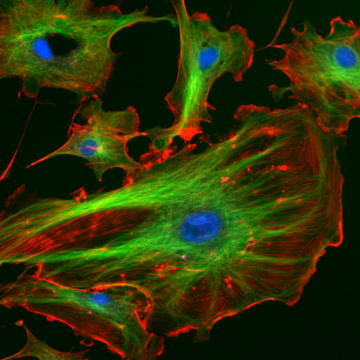
English: a
This image is made from a Molecular Probes demo slide:
Deutsch: Endothelzellen aus der Inneren Wand (Endothel) von Lungenarterien des Rindes unter dem Mikroskop. Die Zellkerne sind mit DAPI blau markiert. Die Mikrotubuli wurden über einen Antikörper grün markiert. Mit rot fluoreszierendem Phalloidin wurden die Aktinfilamente markiert.
Français : Cellulles endothéliales vues au microscope. En bleu, noyaux marqués au DAPI. En vert, microtubules marqués par un anticorps. En rouge, actine marquée à la phalloïdine.
Magyar: Fluoreszcenciamikroszkópos felvétel marha tüdőartéria endotélsejtjeiről (Molecular Probes FluoCells prepared slide #2 (F14781)). A sejtmagok DAPI-val vannak festve (kék), a mikrotubulusokhoz anti-α-tubulin egéranitest, ahhoz pedig BODIPY FL-el jelölt anti-egér kecske-IgG van kapcsolva (zöld), míg az aktin filamentumok Texas Red-X-el kapcsolt falloidinnal vannak jelölve (vörös). A kép három felvétel szuperpozíciójával készült. Hamis színek.
Lietuvių: Citoskeletas. Aktino filamentai – raudona, mikrovamzdeliai – žalia, branduolys – mėlyna spalva.
Română: Sub microscop Celule endoteliale . microtubulii sunt de culoare verde, iar filamentele de actină sunt roşii, pe când nucleul celulei este colorat albastru
Русский: Цитоскелет эукариот. Актиновые микрофиламенты окрашены в красный (фаллоидином, связанным с TRITC), микротрубочки — в зеленый (антителами, связанными с FITC), ядра клеток — в голубой цвет (DAPI). Клетки эндотелия лёгочной артерии быка.
Українська: Цитоскелет еукаріот. Актинові мікрофіламенти забарвлені в червоний колір, мікротрубочки — в зелений, ядра кліток — в блакитний |
| 來源 | http://rsb.info.nih.gov/ij/images/ |
| 作者 | |
| 授權許可 (重用此檔案) |
example image from the ImageJ-Programmpaket (public domain) |
Original file
This image has been taken from the German Wikipedia
The original uploader is de:Benutzer:Jan R. The original upload was at 4th December 2005.
Original description
This image is made from a Molecular Probes demo slide:
Cells: bovine pulmonary arthery endothelial cells
Blue: nucleus stained with DAPI
Green: Tubulin stained with Bodipy FL goat anti-mouse IgG
Red: F-Actin stained with Texas Red X-Phalloidin
(description from [1])
Quelle: Beispielsbild aus dem ImageJ-Programmpaket (public domain), siehe http://rsb.info.nih.gov/ij/
授權條款
此作品在美國屬於公有領域,因為其是由美國政府的官員或僱員,基於其個人公務目的製作的作品,參考美國法典第17篇第1章第105條。
注意︰本模板僅適用於美國聯邦政府的原創作品,不適用於任何美國州、屬地、聯邦個體、縣、市或任何次級政府的作品。本模板也不適用於1978年以後由美國郵政署出版的郵票圖案(參看美國版權局實踐綱領第313.6(C)(1)條)。也不適用於部分美國硬幣;參看美國鑄幣局使用條款。 |
 | |
| 此作品無已知的著作權限制,亦不受所有相關和鄰接的權利限制。 | ||
檔案歷史
點選日期/時間以檢視該時間的檔案版本。
| 日期/時間 | 縮圖 | 尺寸 | 使用者 | 備註 | |
|---|---|---|---|---|---|
| 目前 | 2006年3月24日 (五) 15:07 | | 512 × 512(56 KB) | Splette | {{Information |Description = Endothelial cells under the microscope. Nuclei are stained blue with DAPI, microtubles are marked green by an antibody and actin filaments are labelled red with phalloidin. |Source = http://rsb.info.nih.gov/ij |Date = |Author |
檔案用途
下列9個頁面有用到此檔案:
全域檔案使用狀況
以下其他 wiki 使用了這個檔案:
- af.wikipedia.org 的使用狀況
- ar.wikipedia.org 的使用狀況
- ast.wikipedia.org 的使用狀況
- az.wikipedia.org 的使用狀況
- be.wikipedia.org 的使用狀況
- bg.wikipedia.org 的使用狀況
- bn.wikipedia.org 的使用狀況
- bs.wikipedia.org 的使用狀況
- ca.wikipedia.org 的使用狀況
- ckb.wikipedia.org 的使用狀況
- cs.wikipedia.org 的使用狀況
- cy.wikipedia.org 的使用狀況
- da.wikipedia.org 的使用狀況
- de.wikipedia.org 的使用狀況
- Ultraviolettstrahlung
- Mikrotubulus
- Skelett
- Cytoskelett
- Aktin
- 4′,6-Diamidin-2-phenylindol
- Fluoreszenzmikroskopie
- Listeriose
- Fluoreszenzmarkierung
- Wikipedia Diskussion:Hauptseite/Artikel des Tages/Archiv/Vorschläge/2018/Q3
- Wikipedia:Hauptseite/Archiv/5. August 2018
- Wikipedia Diskussion:Hauptseite/Artikel des Tages/Archiv/Vorschläge/2019/Q1
- Wikipedia:Hauptseite/Archiv/23. März 2019
- de.wikibooks.org 的使用狀況
- de.wikiversity.org 的使用狀況
- en.wikipedia.org 的使用狀況
檢視此檔案的更多全域使用狀況。