File:SDSPAGE.png
SDSPAGE.png (417 × 362像素,文件大小:64 KB,MIME类型:image/png)
From English Wikipedia: http://en.wikipedia.org/w/index.php?title=Image:SDSPAGE.png&action=edit
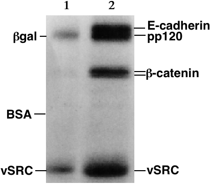
Example of SDS-PAGE of proteins visualized by autoradiography. Two radioactively labeled protein samples were run in adjacent lanes of the gel (1, 2). The larger proteins (β-galactosidase size standard, marker, E-cadherin cell-to-cell adhesion protein, pp120) are towards the top of the gel and smaller proteins (vSRC tyrosine-specific protein kinase, 60,000 Da) are towards the bottom. As its name implies, pp120 is a 120,000 Da phosphoprotein. The β-galactosidase and bovine serum albumin (BSA) size standards were in an adjacent lane (not shown). The radioactive label was 32Phosphate from the gamma position phosphate group of ATP. The vSRC protein is an oncogene that disrupts cell growth by its phosphorylation of other proteins such as β-catenin, a protein that links E-cadherin to the cell's cytoskeleton. In this experiment, the vSRC protein auto-phosphorylated itself and the other proteins (E-cadherin, pp120 and β-catenin). After electrophoresis, medical X-ray film was exposed to the dried gel and regions of dark exposure of the film (the "bands") indicate the position of the radioactively-labeled proteins. Lane 1 is a negative control for which no vSRC was added to the labeling reaction. The other proteins (E-cadherin, pp120 and β-catenin) came from an immunoprecipitation of E-cadherin with anti-E-cadherin antibody. The pp120 and β-catenin proteins exist in a molecular complex with E-cadherin at the surface of the cell and they co-precipitate with E-cadherin. Some cSRC kinase probably also co-precipitated with the E-cadherin, accounting for the faint bands in lane 1. The vSRC kinase was immunoprecipitated from mouse NIH-3T3 cells that had been genetically engineered to express this chicken-derived oncogene. The E-cadherin was from mouse P19 embryonal carcinoma cells. (this picture was worth 290 words)
Uploaded for use on the Gel electrophoresis page.
Source: my personal image.
The copyright to this image is retained by John Schmidt (JWSchmidt).
Permission is granted to copy, distribute and/or modify this image under the terms of the Wikipedia GFDL, as indicated in the fine print at the bottom of this page.
| 本文件采用知识共享署名-相同方式共享 3.0 未本地化版本许可协议授权。 受免責聲明的約束。 | ||
| 署名: JWSchmidt 位于英语维基百科 | ||
| ||
| 本许可协议标签作为GFDL许可协议更新的组成部分被添加至本文件。 |

|
已授权您依据自由软件基金会发行的无固定段落及封面封底文字(Invariant Sections, Front-Cover Texts, and Back-Cover Texts)的GNU自由文件许可协议1.2版或任意后续版本的条款,复制、传播和/或修改本文件。该协议的副本请见“GNU Free Documentation License”。 受免責聲明的約束。 |
If you do not want to use this image under the terms of the GFDL, you can alternatively use it under the terms of the cc-by-nc-sa license.
文件历史
点击某个日期/时间查看对应时刻的文件。
| 日期/时间 | 缩略图 | 大小 | 用户 | 备注 | |
|---|---|---|---|---|---|
| 当前 | 2006年1月1日 (日) 15:10 | | 417 × 362(64 KB) | Llull~commonswiki | From English Wikipedia: http://en.wikipedia.org/w/index.php?title=Image:SDSPAGE.png&action=edit Example of SDS-PAGE of proteins visualized by autoradiography. Two radioactively labeled protein samples were run in adjacent lanes of the gel (1, 2). The la |
文件用途
以下4个页面使用本文件:
全域文件用途
以下其他wiki使用此文件:
- ca.wikipedia.org上的用途
- en.wikipedia.org上的用途
- en.wikibooks.org上的用途
- es.wikipedia.org上的用途
- gl.wikipedia.org上的用途
- ja.wikipedia.org上的用途
- ms.wikipedia.org上的用途