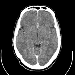
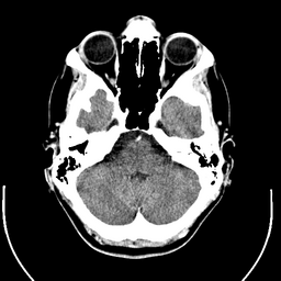
File:Computed tomography of human brain - large.png

預覽大小:800 × 570 像素。 其他解析度:320 × 228 像素 | 640 × 456 像素 | 1,024 × 730 像素 | 1,280 × 913 像素 | 2,560 × 1,826 像素 | 3,639 × 2,595 像素。
原始檔案 (3,639 × 2,595 像素,檔案大小:3.9 MB,MIME 類型:image/png)
檔案歷史
點選日期/時間以檢視該時間的檔案版本。
| 日期/時間 | 縮圖 | 尺寸 | 用戶 | 備註 | |
|---|---|---|---|---|---|
| 目前 | 2017年12月24日 (日) 01:11 |  | 3,639 × 2,595(3.9 MB) | Shashi. | Reverted to version as of 12:49, 1 February 2008 (UTC) |
| 2008年5月8日 (四) 10:59 |  | 3,639 × 2,595(3.17 MB) | CountingPine | Optimise using PNGOUT | |
| 2008年2月1日 (五) 12:49 |  | 3,639 × 2,595(3.9 MB) | Mikael Häggström | {{34 computer tomography images}} {{Individual images of CT of Mikael Häggström's brain}} | |
| 2008年1月31日 (四) 11:56 |  | 3,639 × 2,595(4.03 MB) | Mikael Häggström | {{34 computer tomography images}} {{Individual images of CT of Mikael Häggström's brain}} |
檔案用途
下列8個頁面有用到此檔案:
全域檔案使用狀況
以下其他 wiki 使用了這個檔案:
- bn.wikipedia.org 的使用狀況
- bo.wikipedia.org 的使用狀況
- ca.wikipedia.org 的使用狀況
- en.wikipedia.org 的使用狀況
- CT scan
- Portal:Medicine
- Portal:Medicine/Selected picture
- Portal:Medicine/Selected picture archive
- Wikipedia:WikiProject Neuroscience
- Wikipedia:Featured pictures/Sciences/Biology
- User:Mikael Häggström
- User talk:Mikael Häggström/Archive 1
- Wikipedia:Featured pictures thumbs/10
- Wikipedia:Featured picture candidates/CT of brain of Mikael Häggström.png
- Wikipedia:Featured picture candidates/February-2008
- Wikipedia:Wikipedia Signpost/2008-02-11/Features and admins
- Portal:Medicine/Selected picture/9, 2008
- Portal:Medicine/Selected picture/9
- Wikipedia:Picture of the day/July 2008
- Template:POTD/2008-07-11
- Wikipedia:Wikipedia Signpost/2008-02-11/SPV
- User:Mikael Häggström/Gallery
- Wikipedia:WikiProject Medicine/Recognized content
- Computed tomography of the head
- Wikipedia:Wikipedia Signpost/2013-10-02/Op-ed
- Wikipedia:Wikipedia Signpost/Single/2013-10-02
- User:Wouterstomp/test
- User:Fitness queen04/sandbox
- Wikipedia:WikiProject Anatomy/Resources
- Wikipedia:WikiProject Anatomy/Recognized content
- Wikipedia talk:WikiProject Anatomy/Archive 9
- Reconstruction from projections
- User:VGrigas (WMF)/Quality Media
- User:Flyer22 Frozen/Human brain
- Portal:Medicine/Recognized content
- User talk:Rhododendrites/Reconsidering FPC on the English Wikipedia
- es.wikipedia.org 的使用狀況
- fi.wikipedia.org 的使用狀況
- he.wikipedia.org 的使用狀況
- hy.wikipedia.org 的使用狀況
- hyw.wikipedia.org 的使用狀況
- id.wikipedia.org 的使用狀況
- is.wikipedia.org 的使用狀況
- ja.wikipedia.org 的使用狀況
檢視此檔案的更多全域使用狀況。


