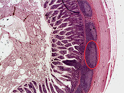
派亞氏淋巴叢
派亞氏淋巴叢(英語:Peyer's patches),又稱培氏斑、派亚氏淋巴丛,是有組織的淋巴結,以17世紀的瑞士解剖學家約翰·康拉德·派亞命名[1]。它們是腸相關淋巴組織的一個重要部分,通常在人類的小腸最低部分發現,主要在遠端空腸和迴腸,但也可以在十二指腸中檢測到[2]。
| 派亞氏淋巴叢 | |
|---|---|
 迴腸的橫切面,派亞氏淋巴叢為圈起部分 | |
| 基本信息 | |
| 系統 | 淋巴系統 |
| 标识字符 | |
| 拉丁文 | noduli lymphoidei aggregati |
| MeSH | D010581 |
| TA98 | A05.6.01.014、A05.7.02.009 |
| TA2 | 2960、2978 |
| TH | H3.04.03.0.00020 |
| FMA | FMA:15054 |
| 《解剖學術語》 [在维基数据上编辑] | |
歷史
编辑派亞氏淋巴叢在17世紀已經被幾個解剖學家觀察和描述過[3],但在1677年,瑞士解剖學家約翰·康拉德·派亞非常清楚地描述這種結狀構造,以至於它們最終以他的名字命名[1][4]。然而派亞認為它們是向小腸排放一些促進消化的物質的腺體。直到1850年,瑞士醫生魯道夫·奧斯卡·齊格勒(Rudolph Oskar Ziegler)在仔細的顯微鏡檢查後提出,派亞氏淋巴叢實際上是淋巴腺[5]。
構造
编辑派亞氏淋巴叢是可觀察到的腸道上皮細胞的拉長增厚,長度為幾公分,在人體中發現約100個。在顯微鏡下,派亞氏淋巴叢顯示為橢圓形或圓形的淋巴濾泡(類似於淋巴結),位於迴腸的黏膜層,並延伸到黏膜下層。派亞氏淋巴叢的數量在15至25歲達到高峰,然後在成年期下降[2]。在迴腸遠端,它們數量眾多,形成一個淋巴環。在人類中至少有46%的派亞氏淋巴叢集中在迴腸遠端25公分處。值得注意的是,不同個體的派亞氏淋巴叢的大小、形狀和分佈都有很大的差異[6]。在成年人中,B細胞被認為是卵泡生髮中心的主導。T細胞存在於濾泡之間的區域。在單核細胞中,CD4+/CD25+(10%)細胞和CD8+/CD25+(5%)細胞在派亞氏淋巴叢中比外周血中更豐富[7]。
派亞氏淋巴叢的特點是濾泡相關上皮(FAE),它覆蓋所有的淋巴濾泡[8]。FAE與典型的小腸絨毛上皮不同:它有較少的杯狀細胞[9] ,因此黏液層較薄[10],而且它的特點是存在專門的M細胞或微皺褶細胞,它們提供從腔內吸收和運輸抗原的功能[8]。此外,與腸道絨毛相比,FAE的基底層更加多孔[11]。最後,FAE細胞對離子和大分子的滲透性較低,這基本上是由於緊密連接蛋白的較高表達[12]。
功能
编辑由於消化道的管腔暴露在外部環境中,其中大部分是潛在的病原微生物,因此派亞氏淋巴叢在腸腔的免疫監視和促進黏膜內免疫反應的產生方面確立了其重要性。
進入腸道的病原微生物和其他抗原會遇到巨噬細胞、樹突狀細胞、B細胞和T細胞,這些細胞在派亞氏淋巴叢和腸相關淋巴組織的其他部位發現。因此派亞氏淋巴叢對消化系統的作用就像扁桃體對呼吸系統的作用一樣,捕獲外來顆粒,監視它們,並將其消滅。
派亞氏淋巴叢被一種特殊的FAE所覆蓋,該上皮含有被稱為微皺褶細胞(M細胞)的特殊細胞,該細胞直接從管腔中取樣抗原並將其傳遞給抗原呈遞細胞(位於其基底面的獨特口袋狀結構中)。樹突狀細胞和巨噬細胞也可以通過將樹突延伸到經細胞的M細胞特異性孔中來直接對腔體進行採樣[13][14]。同時FAE細胞的旁路被緊緊關閉,以防止抗原的滲透和與免疫細胞的持續接觸[15]。T細胞、B細胞和記憶細胞在派亞氏淋巴叢遇到抗原時受到刺激,然後這些細胞傳到腸系膜淋巴結,免疫反應在那裡被放大。被激活的淋巴細胞通過胸導管進入血流,並前往腸道,在那裡執行其最終的效應器功能。 B細胞的成熟是在派亞氏淋巴叢中進行的。
臨床意義
编辑儘管在免疫反應中很重要,但派亞氏淋巴叢中淋巴組織的過度生長是病理性的,因為派亞氏淋巴叢的肥大與特發性腸套疊密切相關。
擁有過多或大於正常的派亞氏淋巴叢與朊毒體疾病的風險增加有關。
參見
编辑參考資料
编辑- ^ 1.0 1.1 Peyer, Johann Conrad. Exercitatio Anatomico-Medica de Glandulis Intestinorum, Earumque Usu et Affectionibus [Anatomical-medical essay on the intestinal glands, and their function and diseases]. Schaffhausen, Switzerland: Onophrius à Waldkirch. 1677 (Latin).
- Reprinted as: Peyer, Johann Conrad. Exercitatio Anatomico-Medica de Glandulis Intestinorum, Earumque Usu et Affectionibus. Amsterdam, Netherlands: Henrik Wetstein. 1681 [2022-10-02]. (原始内容存档于2022-10-02) (Latin).
- Peyer referred to Peyer's patches as plexus or agmina glandularum (clusters of glands). From (Peyer, 1681), p. 7: "Tenui a perfectiorum animalium Intestina accuratius perlustranti, crebra hinc inde, variis intervallis, corpusculorum glandulosorum Agmina sive Plexus se produnt, diversae Magnitudinis atque Figurae." (I knew from careful study of more advanced animals, the intestines bear — often here and there, at various intervals — clusters of glandular small bodies or "plexuses" of diverse size and shape.) From p. 15: "(has Plexus seu agmina Glandularum voco)" (I call them "plexuses" or clusters of glands) He described their appearance. From p. 8: "Horum vero Plexuum facies modo in orbem concinnata; modo in Ovi aut Olivae oblongam, aliamve angulosam ac magis anomalam disposita figuram cernitur." (But the configurations of these "plexuses" are arranged at one time in a circle; at another time, it is seen in an egg [shape] or an oblong olive [shape] or other faceted and more irregularly arranged shape.) Drawings of Peyer's patches appear after pages 22 and 24.
- ^ 2.0 2.1 Zijlstra M, Auchincloss H, Loring JM, Chase CM, Russell PS, Jaenisch R. Skin graft rejection by beta 2-microglobulin-deficient mice. The Journal of Experimental Medicine. April 1992, 175 (4): 885–93. PMC 1552287 . PMID 18668776. doi:10.1136/gut.6.3.225.
- ^ Haller, Albrecht von. Elementa Physiologiae corporis humani [Elements of the physiology of the human body] 7. Bern, Switzerland: Societas Typographica. 1765: 35 [2022-10-02]. (原始内容存档于2022-10-04) (Latin). Anatomists who mentioned Peyer's patches included:
- Johann Theodor Schenck (1619 – 1671): Schenck, Johann Theodor. Exercitationes Anatomicæ ad Usum Medicum Accommodatæ [Anatomical Exercises Suited to Medical Practice]. Jena, (Germany): Johann Ludwig Neuenhahn. 1672: 334 [2022-10-02]. (原始内容存档于2022-10-07) (Latin). Schenk thought that intestinal worms resided in Peyer's patches and that the orifices of the patches were the worms' mouths. From p. 334: "In canibus saepissime observavi non ad ventriculum … a praeter labente chylo sibi conveniens allicerent." (In dogs, I very often noticed — not only near the stomach but also on the walls of their small intestines — flesh-colored or glandular blisters, [appearing] to swim one after another, [in] which, when we dissected [them], I observed some smooth reddish worms [vermium] living there in clusters [with] their heads facing towards the cavity of the intestines, in which part there were glands with orifices, [but] reversed, so that from there they obtained, from the chyle flowing past, nourishment [that was] suitable for them.)
- Jeremias Loss (1643 – 1684): Loss, Jeremias. Dissertatio Medica de Glandulis in Genere [Medical Discourse on Glands in [Various] Species]. Wittenberg, (Germany): Martin Schultz. 1683: 12 [2022-10-02]. (原始内容存档于2022-10-07) (Latin). On page 12, Loss states that some glands are located "inter Membranas viscerum quorundam" (between the membranes of certain internal organs) " … prout id in Glandulis Intestinorum satis manifestum est." (as it is quite clear in the glands of the intestines), where "In Intestinis ita congregantur, interdum pauciores, interdum plures, ut areolas quasdam constituant: … " (in the intestines there are thus gathered sometimes fewer [glands], sometimes more [glands], so that they form certain round patches.)
- Johannes Nicolaus Pechlin (1646 – 1706): Pechlin, Johannes Nicolaus. De Purgantium Medicamentorum Facultatibus [On the Means of Medicinal Purges]. Leiden and Amsterdam, Netherlands: Daniel, Abraham, and Adrian à Gaasbeek. 1672: 510 [2022-10-02]. (原始内容存档于2022-10-02) (Latin). From p. 510: " … ego tenuium glandularum glomeratum agmen esse ratus, … " (… I considered the heaped cluster of fine glands, … )
- Martin Lister (ca. 1638 – 1712): Lister, Martin. A letter of Mr Lister dated May 21. 1673. in York, partly taking notice of the foregoing intimations, partly communicating some anatomical observations and experiments concerning the unalterable character of the whiteness of the chyle within the lacteous veins; together with divers particulars observed in the guts, especially several sorts of worms found in them.. Philosophical Transactions of the Royal Society of London. 23 June 1673, 8 (95): 6060–6065 [2022-10-02]. Bibcode:1673RSPT....8.6060L. doi:10.1098/rstl.1673.0026. (原始内容存档于2022-10-06). From p. 6062: "As 1. Glandulae miliares of the small Guts, which may also in some Animals be well call'd fragi-formes, from the figure of the one half of a Strawberry, and which yet I take to be Excretive glanduls, because Conglomerate."
- Nehemiah Grew (1641 – 1712): Grew, Nehemiah. The Comparative Anatomy of Stomachs and Guts Begun. Being Several Lectures Read before the Royal Society. In the Year, 1676.. London, England: Self-published. 1681: 3 [2022-10-02]. (原始内容存档于2022-10-02). Grew called Peyer's patches pancreas intestinale.
- ^ There were many earlier names for Peyer's patches:
- Todd, Robert Bentley (编). The Cyclopædia of Anatomy and Physiology 5. London, England: Longman, Brown, Green, Longmans, & Roberts. 1859: 356 footnote [2022-10-02]. (原始内容存档于2022-10-07).
- Leidy, Joseph. An Elementary Treatise on Human Anatomy. Philadelphia, Pennsylvania, USA: J.B. Lippincott & Co. 1861: 313 footnote [2022-10-02]. (原始内容存档于2022-10-06).
- ^ Ziegler, Rudolph Oskar (1850) Ueber die solitären und Peyerschen Follikel : Inaugural-Abhandlung, der medicinischen Facultät der Julius-Maximilians-Universität zu Würzburg vorgelegt [On solitary and Peyer's follicles: Inaugural treatise, submitted to the medical faculty of the Julius-Maximilians-University of Würzburg] (in German) Würzburg, (Germany): Friederich Ernst Thein. From p. 37: "Ebensogross, wo nicht grösser ist die Aehnlichkeit der sogenannten Peyer'schen Drüsen und der Lymphdrüsen." (Just as great, if not greater, is the resemblance between the so-called Peyer's glands and the lymph glands.) From p. 38: " … ja, man könnte selbst versucht sein, die letzteren für nichts als eine Art von zwischen den Wänden der Darmsschleimhaut eingebetteten Lymphdrüsen zu halten." ( … indeed, one could even be tempted to regard the latter [i.e., the Peyer's patches] as nothing but some type of lymph glands [which are] embedded between the walls of the intestinal mucosa.)
- ^ Van Kruiningen HJ, West AB, Freda BJ, Holmes KA. Distribution of Peyer's patches in the distal ileum. Inflammatory Bowel Diseases. May 2002, 8 (3): 180–5. PMID 11979138. S2CID 22514793. doi:10.1097/00054725-200205000-00004.
- ^ Jung C, Hugot JP, Barreau F. Peyer's Patches: The Immune Sensors of the Intestine. International Journal of Inflammation. September 2010, 2010: 823710. PMC 3004000 . PMID 21188221. doi:10.4061/2010/823710.
- ^ 8.0 8.1 Owen RL, Jones AL. Epithelial cell specialization within human Peyer's patches: an ultrastructural study of intestinal lymphoid follicles. Gastroenterology. February 1974, 66 (2): 189–203. PMID 4810912. doi:10.1016/s0016-5085(74)80102-2 .
- ^ Onori P, Franchitto A, Sferra R, Vetuschi A, Gaudio E. Peyer's patches epithelium in the rat: a morphological, immunohistochemical, and morphometrical study. Digestive Diseases and Sciences. May 2001, 46 (5): 1095–104. PMID 11341655. S2CID 34204173. doi:10.1023/a:1010778532240.
- ^ Ermund A, Gustafsson JK, Hansson GC, Keita AV. Mucus properties and goblet cell quantification in mouse, rat and human ileal Peyer's patches. PLOS ONE. 2013, 8 (12): e83688. Bibcode:2013PLoSO...883688E. PMC 3865249 . PMID 24358305. doi:10.1371/journal.pone.0083688 .
- ^ Takeuchi T, Gonda T. Distribution of the pores of epithelial basement membrane in the rat small intestine. The Journal of Veterinary Medical Science. June 2004, 66 (6): 695–700. PMID 15240945. doi:10.1292/jvms.66.695 .
- ^ Markov AG, Falchuk EL, Kruglova NM, Radloff J, Amasheh S. Claudin expression in follicle-associated epithelium of rat Peyer's patches defines a major restriction of the paracellular pathway. Acta Physiologica. January 2016, 216 (1): 112–9. PMID 26228735. S2CID 13389571. doi:10.1111/apha.12559. hdl:11701/6438 .
- ^ Lelouard H, Fallet M, de Bovis B, Méresse S, Gorvel JP. Peyer's patch dendritic cells sample antigens by extending dendrites through M cell-specific transcellular pores. Gastroenterology. March 2012, 142 (3): 592–601.e3. PMID 22155637. doi:10.1053/j.gastro.2011.11.039.
- ^ Bonnardel J, Da Silva C, Henri S, Tamoutounour S, Chasson L, Montañana-Sanchis F, Gorvel JP, Lelouard H. Innate and adaptive immune functions of peyer's patch monocyte-derived cells. Cell Reports. May 2015, 11 (5): 770–84. PMID 25921539. doi:10.1016/j.celrep.2015.03.067 .
- ^ Diener M. Roadblock for antigens--take a detour via M cells. Acta Physiologica. January 2016, 216 (1): 13–4. PMID 26335934. doi:10.1111/apha.12595 .
- ^ Pascall, C R; Stearn, E J; Mosley, J G, Short Reports, British Medical Journal 281 (6232), 1980-07-05: 26, PMC 1713722 , PMID 7407483, doi:10.1136/bmj.281.6232.26-a,
Unlike S hadar peritonitis, S typhi peritonitis is due to perforation of Peyer's patches.