囊泡
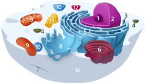
囊泡(英语:vesicle)又称小泡,在细胞生物学中指一类细胞内体积相对较小,有特殊内含物的封闭囊状构造,其外围由至少一层的脂质双层分子膜(单位膜)包围而成,用来存放、消化、转运物质(例如细胞产物或废物)。
| 细胞生物学 | |
|---|---|
| 动物细胞 | |

|
分类
编辑液泡
编辑液泡(vacuole)是主要含有水的一种细胞器,由多囊体的融合衍生而成[1]。植物细胞在细胞的中心部分有一个较大的液泡,用于控制渗透和营养物质的储存。在某些生物中,特别是在纤毛虫中发现了具有收缩性的液泡。 这些液泡从细胞质中吸收水分,并将其从细胞中排出,以避免由于渗透压而破裂。
溶酶体
编辑溶酶体(lysosome)外包单层膜,膜上有转运蛋白,而内部则富含酸性水解酶,负责吞噬及自噬等降解途径将进入细胞的外源性大分子物质和出现异常的大分子物质降解,从而为细胞提供氨基酸及脂质等营养物质[2],并且参与质膜修复及细胞信号传导等细胞过程。
脂质体
编辑脂质体(liposome)是磷脂分散在水中形成的一个包封着部分水相(aqueous phase)的类球状封闭囊泡。脂质体的形态与细胞膜相似,并且能够融合各种物质。脂质体被认为是体外和体内生物活性物质的最佳递送系统,以及最成功的药物载体系统。
运输囊泡
编辑运输囊泡(transport vesicles)在真核生物中,通过在不同细胞器及细胞表面进行转运发挥它的作用。目前已知的运输囊泡有网格蛋白囊泡、外被体蛋白Ⅰ(coat protein、COPI囊泡)和COPⅡ囊泡。这些运输囊泡在细胞膜的生物合成、细胞器功能和形态的动态平衡,以及蛋白质分泌等方面都发挥重要作用[3]。目前已知在真核生物中,约有三分之一蛋白质的早期分泌途径依赖着COPⅡ囊泡的运输。早期分泌途径中,COPⅡ囊泡介导新合成的分泌性蛋白质和膜脂质等货物从内质网转运至顺面高尔基复合体(Golgi)。COPⅠ囊泡则介导货物分子从顺面Golgi逆向转运至内质网,以及货物分子在Golgi膜囊间的转运[4][5]。晚期分泌途径中,含衔接蛋白-1(adaptor protein 1、AP-1)、AP-3及AP-4蛋白复合物的网格蛋白囊泡可参与货物分子在反面Golgi与内体、溶酶体及质膜间的转运[6][7]。含AP-2蛋白的网格蛋白囊泡参与货物的胞吞途径,通过质膜内陷形成囊泡,将细胞外或质膜表面的货物包裹到囊泡内,并且运入细胞[8]。
- COPⅠ囊泡:最初研究者利用三磷酸鸟苷(GTP)衍生物GTPγS(一种富含高尔基体膜的细胞质与抗水解的GTP衍生物)共培养时,发现高尔基体池之间存在一种囊泡转运结构[9](后来在真核细胞中也证实此结构的存在[10])。除了脂质成分外,参与此囊泡形成的成分还有7种外被体蛋白(即外被体α、β、β′、γ、δ、ε、ζ)。这些外被体蛋白相互作用形成的复合物就是COPⅠ囊泡[11][12]。亚单位α、β′、ε在结构上与网格蛋白及COPⅡ囊泡的外层组分具有较高的一致性,形成复合物的内层组分称为B亚复合物(主要负责与靶蛋白结合),而亚单位β、γ、δ、ζ 与网格蛋白及COPⅡ囊泡的内层组分相似,形成复合物的内层组分称为F亚复合物,该亚复合物主要负责与靶蛋白结合,并且直接与COPⅠ囊泡形成的招募者ADP核糖基化因子(ADP ribosylation factors)结合,从而参与启动囊泡的逆向转运[11][12][13]。同时又发现COPγ、COPζ各有2种亚型,即γ1、γ2、ζ1、ζ2[14],这些亚型间的交叉组合可形成4种COPⅠ囊泡。有研究发现,3T3细胞中约五成的COPⅠ囊泡包含γ1ζ1,约三成包含γ2ζ1,约两成包含γ1ζ2,仅有不足半成的COPⅠ囊泡包含γ2ζ2[15]。另外一份研究则发现细胞中,超过七成的γ1型COPⅠ复合物定位于顺式高尔基体,超过六成的γ2型COPⅠ复合物定位于反式高尔基体,约八成的ζ1型COPⅠ复合物定位于顺式高尔基体,而ζ2型COPⅠ复合物因缺乏特异性抗体而尚未明确定位[16]。由此可见,COPⅠ囊泡的类型,可能与囊泡的起源部位,以及定向转运至的靶细胞器膜有一定的关系[17]。
- COPⅡ囊泡:主要是由五种可溶性蛋白,即小GTP酶蛋白Sar1、Sec23及Sec24(这两种蛋白会紧密结合形成蝴蝶结状异二聚体)、Sec13及Sec31组成的复合物[18],并且以分层方式按顺序组装至内质网膜上,并且介导COPⅡ囊泡的形成。根据COPⅡ囊泡装配的货物分子大小,Sec31 C端的氨基酸序列可以成不同几何形状的COPⅡ笼型,从而使COPⅡ囊泡容纳前胶原、乳糜微粒及载脂蛋白等超过COPⅡ囊泡本身大小的大型货物分子[19],这可能与E3泛素连接酶通过其配体衔接蛋白KLHL12,使Sec31泛素化有关[20][21],从而令Sec31招募到有利于形成管状COPⅡ衣被结构的其他因子,同时调节Sec23的活性而激活Sar1-GTP的GTP酶水解活性。除了上述五种可溶性蛋白外,进一步研究发现Sec12和Sec16对COPⅡ囊泡的形成也非常重要[22]。
分泌泡
编辑分泌泡(secretory vesicle)包含将要从细胞中排出的物质。细胞排泄物质的原因有很多,其中一个原因是要处理废物,而另一个原因与细胞的功能有关。在较大的生物体内,某些细胞专门产生某些化学物质,而这些化学物质储存在分泌泡中,并且在需要时释放出来。
- 膜囊泡:细菌、古菌、真菌及寄生虫会释放出膜囊泡(membrane vesicles),而这些膜囊泡含有不同但专门的有毒化合物和生化信号分子,这些膜囊泡会被转运到靶细胞,以启动有利于微生物的过程(例如入侵宿主细胞和杀死在竞争性微生物)[23]。
细胞外囊泡
编辑细胞外囊泡(extracellular vesicles,EVs)是一类由活细胞旁分泌释放到周围环境中的双层脂质膜结构的微小囊泡,主要分为外排体(exosome)、微囊泡(microvesicle)即微粒(microparticle)、凋亡小体(apoptosis body)及癌小体(oncosome)四类,然而尚可以细分为核外粒体(ectosome)、神经突触小体(Synaptic vesicle)及基质小泡(matrix vesicle)等,可以由复杂的真核生物,包括革兰氏阴性菌和革兰氏阳性菌、分枝杆菌及真菌等产生[24][25],几乎所有细胞都能够释放EVs。
- 外排体:外定义的变异范围从30~100 nm,40~100 nm,50~150 nm到40~200 nm[26][27][28][29],然而亦有指其直径通常在30~100 nm,具有双层分子膜结构,形态呈球形小体、杯状或扁形,而在体液中主要以球形小体的形式存在,并且可以通过超速离心的方法获得。外排体存在于血清、血浆、尿液、母乳及恶性体液(例如恶性胸水及恶性腹水等),几乎所有类型的细胞都能释放外排体。外排体中包含各种不同的成分,例如蛋白质、酶类、去氧核糖核酸、信使核糖核酸、非编码核糖核酸及脂质等,并且含有细胞因子及生长因子受体等。外排体被认为是细胞间通讯的第3条途径,积极地充当不同组织和细胞之间的生物活性分子。此外,外排体广泛地参与免疫反应,因为它携带来源细胞的细胞膜表面相关抗原、生物活性小分子物质、免疫刺激因子及免疫抑制分子等。除此之外,外排体可以调节肿瘤微环境,因为肿瘤细胞释放的外泌体有利于非恶性组织的恶化,从而促进肿瘤转移。
- 微囊泡、微粒:是机体各种细胞在正常或病理状态下从细胞质膜上脱落而释放的膜性小囊泡,直径通常在30至1 000 nm。血小板在正常生理状态下是微囊泡的来源[30],当内皮细胞及血液细胞[31]等细胞发生激活或凋亡时,在细胞因子及剪切力等触发因素下也可以产生微囊泡。首先,微囊泡的表面配体与靶细胞的受体结合,激活或者抑制靶细胞内的转导通路,例如促进组织因子介导的凝血反应。其次,微囊泡形成时会包裹母细胞的成分,并且传递到靶细胞中,使其具有新的生物学功能。第三,微囊泡会将母细胞的受体转移到靶细胞而呈现相关受体表型。第四,微囊泡能转移完整的细胞器或病毒等致病因子,并且传递给其他细胞[32][33][34]。
- 癌小体:
- 核外粒体:一种在质膜处聚集并从质膜释放的细胞外囊泡,其标记物有TyA和C1a(而非外排体的标记物CD63和CD61)。尽管英文名称与外排体相似,但是并非同一种细胞外囊泡。因为外排体从多囊体释放,而核外粒体则被快速组装在质膜中,在特定的细胞刺激下会大大增加,最终从细胞的表面释放[35]。此外,外排体在其初始状态下的表现为向着多囊体内含物方向的向内小芽,而核外粒体在初始状态下的表现则为离散的质膜结构域,并且以与其胞质表面相关的致密物质层作为标志物,核外粒体的结构域在几秒或最多几分钟内就会向外发芽并被夹成圆形的囊泡[35]。释放出来的核外粒体和外排体均包含着由蛋白质、RNA和DNA序列组成的基质,然而在核外粒体中的含量更高[35]。目前已知核外粒体拥有着多种英文异称,包括“nanoparticles”、“microparticles”、“microvesicles”、“shedding vesicles”、“shedding bodies”、“exovesicles”、“secretory vesicles”,以及“oncosomes”[35]。
- 基质小泡:基质小泡位于细胞外的空间或细胞外基质内。 1967年,Clarke Anderson及Ermanno Bonucci分别使用电子显微镜发现基质小泡的存在[36][37]。这些源自细胞的基质小泡专门用于启动各种组织(包括骨骼、软骨和牙本质)中基质的生物矿化作用。在正常钙化过程中,钙和磷酸根离子大量流入细胞,伴随着细胞凋亡(从基因上确定为自毁)和基质小泡的形成。钙负荷会导致磷脂酰丝氨酸:钙:磷酸盐复合物在质膜中形成,部分由annexin所介导。基质囊泡从质膜发芽,准确来说是在与细胞外基质相互作用的位点。因此,基质小泡将钙、磷酸盐、脂质和annexin传递到细胞外基质,其作用帮助形成成核矿物质。 除非高尔基体不存在,否则这些过程将被精确地协调,在适当的时间和地点实现组织基质的矿化。
循环细胞膜囊泡
编辑循环细胞膜囊泡(tissue factor-carrying microvesicle,TF-MV)是一种细胞在激活、受损或凋亡时从细胞膜上释放的脂质囊泡(直径约0.1~1 μm),具有促凝活性的TF-MV最主要来源于动脉粥样硬化斑块[38][39]。TF-MV携有母体细胞内容物或特异性表面蛋白,并且因可以结合受体细胞而会影响内皮功能障碍及凝血等疾病的生物学过程。目前已知其在静脉血栓形成及相关疾病的发生及发展中占据重要的作用,可以独立地触发凝血级联反应[40],甚至可以用来预测血栓性疾病的发生和发展,以及严重程度分级[41][42][43][44]。有研究指出参与急性缺血性卒中,血管炎症过程的炎症介质正是循环白细胞膜囊泡,可以促进颅内血管动脉粥样硬化的进程[45],然而组织因子途径抑制物(TFPI)的表达会随着TF-MV的表达增加而明显增加[43],可以称是生物体内凝血与抗凝的激活矛盾[46]。
其他类别
编辑- 气泡(gas vesicle)存于古菌、细菌、浮游生物,可能用于调节浮力,通过调节气体含量来控制垂直迁移,或者可能为了最大程度地收集太阳光而定位细胞。 这些囊泡通常表现为由蛋白质制成的柠檬形或圆柱形管[47] ,它们的直径决定了囊泡的强度,较大的囊泡则较弱。 囊泡的直径也影响其体积及其提供浮力的效率。 蓝菌门细菌已努力制造出拥有最大直径的囊泡,同时保持结构稳定。蛋白质可以让气体穿过,但是不能让水穿过,从而防止囊泡出现“泛滥”[48]。
- 多囊体(multivesicular body,MVB)是一种次级胞内体(late endosome),并且是一种包含着许多小囊泡的膜结合囊泡。
电分析化学
编辑电分析化学法具有时间分辨率和灵敏度均较高的特点,而研究人员先前对胞吐过程进行研究时所采用的光学方法(例如引入光学标记分子及光谱成像等),虽然在研究胞吐过程中发挥着重要作用[49][50][51],但是这些方法均存在着时间分辨率低及定量困难等缺点。因此,能够克服这些缺点的电分析化学法在单神经囊泡的分析方面,拥有着颇大的优势[52]。
1991年, 有科研人员首次采用一种名为“单细胞电流法”的技术,实现胞吐过程中单囊泡释放神经递质的定量、动态、快速分析[53][54]。许多研究人员随后使用这个方法研究药物及环境等因素对囊泡释放神经递质的影响[55][56][57]。
单囊泡原位电化学计数法
编辑目前已知瑞典哥德堡大学的科研人员分成不同研究小组,并且在此方面做出一系列的工作,发展三种电化学计数法,即毛细管电泳-电化学计数法、单囊泡电化学计数法(VIEC),以及单囊泡原位电化学计数法(IVIEC)[52]。
- 毛细管电泳-电化学计数法:这是第一套单囊泡内包被神经递质的定量分析装置,巧妙地结合了毛细管电泳、微流控芯片和电化学检测技术。由于毛细管电泳的毛细管出口被固定在聚二甲基硅氧烷制备的微流控芯片上,而且在出口处引入囊泡膜裂解液(即高浓度十二烷基磺酸钠),以及放置柱状碳纤维微电极。因此,在毛细管内的单个囊泡(已被电场分离出),会被裂解液破开囊泡膜,使电极可以完全检测神经递质,实现单一囊泡储存神经递质的定量测定[58]。成功地令PC12细胞(一种大鼠肾上腺嗜铬细胞瘤细胞)及鼠脑组织分离出的神经囊泡内包被神经递质,可以被定量分析[59][60],然而这种定量分析装置存在复杂且难于操控的缺点[52]。
- 单囊泡电化学计数法:2015年,有科技人员发展了VIEC,而VIEC不需要预先进行电泳,就可以裂解电极表面的单个囊泡,从而定量分析单个囊泡内包被神经递质。在装置的盘状碳电极上,从肾上腺髓质中分离出的肾上腺嗜铬囊泡会发生反应,儿茶酚胺类神经递质等囊泡内包被神经递质,被电极电化学氧化,从而实现检测[61]。其他与之相似的单囊泡电化学计数法在此后陆续被其他研究人员所开发[62][63][64],并且具有一定的研究价值,例如发现囊泡会在电极的表面形成一个很快就消失的纳米小孔[65],而该小孔的位置对电极上检测到释放神经递质的比例有影响[66],又例如发现囊泡膜破裂的驱动力主要是电穿孔[67],而电穿孔的发生会被膜内多肽或膜蛋白的存在所阻止[67]。除此之外,有研究人员发现温度提高后容易诱发电穿孔的形成,因为囊泡膜内的膜蛋白在磷脂双分子层中的运动速率会增大,并且提高膜磷脂与电极紧密结合的可能性[65];另外又有研究人员发现尺寸愈大的囊泡因与电极接触的面积越大,囊泡膜磷脂与电极形成紧密结合的点位越多,而更容易发生电穿孔[65][68]。
参见
编辑参考文献
编辑- ^ Cui, Y; Cao, W; He, Y; Zhao, Q; Wakazaki, M; Zhuang, X; Gao, J; Zeng, Y; Gao, C; Ding, Y; Wong, HY; Wong, WS; Lam, HK; Wang, P; Ueda, T; Rojas-Pierce, M; Toyooka, K; Kang, BH; Jiang, L. A whole-cell electron tomography model of vacuole biogenesis in Arabidopsis root cells.. Nature plants. 2019-01, 5 (1): 95–105 [2020-02-10]. PMID 30559414. doi:10.1038/s41477-018-0328-1. (原始内容存档于2020-03-03).
- ^ Saftig, P; Klumperman, J. Lysosome biogenesis and lysosomal membrane proteins: trafficking meets function.. Nature reviews. Molecular cell biology. 2009-09, 10 (9): 623–35 [2020-02-11]. PMID 19672277. doi:10.1038/nrm2745. (原始内容存档于2020-03-03).
- ^ Béthune, J; Wieland, FT. Assembly of COPI and COPII Vesicular Coat Proteins on Membranes.. Annual review of biophysics. 2018-05-20, 47: 63–83 [2020-02-12]. PMID 29345989. doi:10.1146/annurev-biophys-070317-033259.[永久失效链接]
- ^ Bacia, K. Intracellular transport mechanisms: Nobel Prize for Medicine 2013.. Angewandte Chemie (International ed. in English). 2013-11-25, 52 (48): 12486–8 [2020-02-12]. PMID 24249548. doi:10.1002/anie.201308937. (原始内容存档于2020-03-03).
- ^ Faini, M; Beck, R; Wieland, FT; Briggs, JA. Vesicle coats: structure, function, and general principles of assembly.. Trends in cell biology. 2013-06, 23 (6): 279–88 [2020-02-12]. PMID 23414967. doi:10.1016/j.tcb.2013.01.005.[永久失效链接]
- ^ Robinson, MS. Forty Years of Clathrin-coated Vesicles.. Traffic (Copenhagen, Denmark). 2015-12, 16 (12): 1210–38 [2020-02-12]. PMID 26403691. doi:10.1111/tra.12335. (原始内容存档于2020-03-03).
- ^ Guo, Y; Sirkis, DW; Schekman, R. Protein sorting at the trans-Golgi network.. Annual review of cell and developmental biology. 2014, 30: 169–206 [2020-02-12]. PMID 25150009. doi:10.1146/annurev-cellbio-100913-013012.[永久失效链接]
- ^ Kirchhausen, T; Owen, D; Harrison, SC. Molecular structure, function, and dynamics of clathrin-mediated membrane traffic.. Cold Spring Harbor perspectives in biology. 2014-05-01, 6 (5): a016725 [2020-02-12]. PMID 24789820. doi:10.1101/cshperspect.a016725. (原始内容存档于2020-03-03).
- ^ Malhotra, V; Serafini, T; Orci, L; Shepherd, JC; Rothman, JE. Purification of a novel class of coated vesicles mediating biosynthetic protein transport through the Golgi stack.. Cell. 1989-07-28, 58 (2): 329–36 [2020-02-12]. PMID 2752426. doi:10.1016/0092-8674(89)90847-7.[永久失效链接]
- ^ Waters, MG; Serafini, T; Rothman, JE. 'Coatomer': a cytosolic protein complex containing subunits of non-clathrin-coated Golgi transport vesicles.. Nature. 1991-01-17, 349 (6306): 248–51 [2020-02-12]. PMID 1898986. doi:10.1038/349248a0 (英语).[永久失效链接]
- ^ 11.0 11.1 Hara-Kuge, S; Kuge, O; Orci, L; Amherdt, M; Ravazzola, M; Wieland, FT; Rothman, JE. En bloc incorporation of coatomer subunits during the assembly of COP-coated vesicles.. The Journal of cell biology. 1994-03, 124 (6): 883–92 [2020-02-12]. PMID 8132710. doi:10.1083/jcb.124.6.883. (原始内容存档于2020-03-03).
- ^ 12.0 12.1 Jackson, LP. Structure and mechanism of COPI vesicle biogenesis.. Current opinion in cell biology. 2014-08, 29: 67–73 [2020-02-12]. PMID 24840894. doi:10.1016/j.ceb.2014.04.009. (原始内容存档于2020-03-03).
- ^ Lee, C; Goldberg, J. Structure of coatomer cage proteins and the relationship among COPI, COPII, and clathrin vesicle coats.. Cell. 2010-07-09, 142 (1): 123–32 [2020-02-12]. PMID 20579721. doi:10.1016/j.cell.2010.05.030.[永久失效链接]
- ^ Blagitko, N; Schulz, U; Schinzel, AA; Ropers, HH; Kalscheuer, VM. gamma2-COP, a novel imprinted gene on chromosome 7q32, defines a new imprinting cluster in the human genome.. Human molecular genetics. 1999-12, 8 (13): 2387–96 [2020-02-12]. PMID 10556286. doi:10.1093/hmg/8.13.2387.[永久失效链接]
- ^ Beck, R; Rawet, M; Wieland, FT; Cassel, D. The COPI system: molecular mechanisms and function.. FEBS letters. 2009-09-03, 583 (17): 2701–9 [2020-02-12]. PMID 19631211. doi:10.1016/j.febslet.2009.07.032.[永久失效链接]
- ^ Moelleken, J; Malsam, J; Betts, MJ; Movafeghi, A; Reckmann, I; Meissner, I; Hellwig, A; Russell, RB; Söllner, T; Brügger, B; Wieland, FT. Differential localization of coatomer complex isoforms within the Golgi apparatus.. Proceedings of the National Academy of Sciences of the United States of America. 2007-03-13, 104 (11): 4425–30 [2020-02-12]. PMID 17360540. doi:10.1073/pnas.0611360104.[永久失效链接]
- ^ ZHANGGuang-ya; XIONGJie; CHENFeng-ling. Research Progress in the Structure and Function of Coat Protein Ⅰ. Yixue Zongshu. 2016, (1): 5–9. doi:10.3969/j.issn.1006-2084.2016.01.002.
- ^ Matsuoka, K; Orci, L; Amherdt, M; Bednarek, SY; Hamamoto, S; Schekman, R; Yeung, T. COPII-coated vesicle formation reconstituted with purified coat proteins and chemically defined liposomes.. Cell. 1998-04-17, 93 (2): 263–75 [2020-02-12]. PMID 9568718. doi:10.1016/s0092-8674(00)81577-9.[永久失效链接]
- ^ Saito, K; Katada, T. Mechanisms for exporting large-sized cargoes from the endoplasmic reticulum.. Cellular and molecular life sciences : CMLS. 2015-10, 72 (19): 3709–20 [2020-02-12]. PMID 26082182. doi:10.1007/s00018-015-1952-9.[永久失效链接]
- ^ Lord, C; Ferro-Novick, S; Miller, EA. The highly conserved COPII coat complex sorts cargo from the endoplasmic reticulum and targets it to the golgi.. Cold Spring Harbor perspectives in biology. 2013-02-01, 5 (2) [2020-02-12]. PMID 23378591. doi:10.1101/cshperspect.a013367.[永久失效链接]
- ^ Malhotra, V; Erlmann, P. The pathway of collagen secretion.. Annual review of cell and developmental biology. 2015, 31: 109–24 [2020-02-12]. PMID 26422332. doi:10.1146/annurev-cellbio-100913-013002.[永久失效链接]
- ^ Khoriaty, R; Vasievich, MP; Ginsburg, D. The COPII pathway and hematologic disease.. Blood. 2012-07-05, 120 (1): 31–8. PMID 22586181. doi:10.1182/blood-2012-01-292086.
- ^ Deatherage, B. L.; Cookson, B. T. Membrane Vesicle Release in Bacteria, Eukaryotes, and Archaea: a Conserved yet Underappreciated Aspect of Microbial Life. Infection and Immunity. 2012, 80 (6): 1948–1957. ISSN 0019-9567. PMC 3370574 . PMID 22409932. doi:10.1128/IAI.06014-11.
- ^ Yáñez-Mó M, Siljander PR, Andreu Z, et al. Biological properties of extracellular vesicles and their physiological functions. J Extracell Vesicles. 2015, 4: 27066. PMC 4433489 . PMID 25979354. doi:10.3402/jev.v4.27066.
- ^ Théry C, Witwer KW, Aikawa E, et al. Minimal information for studies of extracellular vesicles 2018 (MISEV2018): a position statement of the International Society for Extracellular Vesicles and update of the MISEV2014 guidelines. J Extracell Vesicles. 2018, 7 (1): 1535750. PMC 6322352 . PMID 30637094. doi:10.1080/20013078.2018.1535750.
- ^ Demory Beckler, M; Higginbotham, JN; Franklin, JL; Ham, AJ; Halvey, PJ; Imasuen, IE; Whitwell, C; Li, M; Liebler, DC; Coffey, RJ. Proteomic analysis of exosomes from mutant KRAS colon cancer cells identifies intercellular transfer of mutant KRAS.. Molecular & cellular proteomics : MCP. 2013-02, 12 (2): 343–55 [2020-02-10]. PMID 23161513. doi:10.1074/mcp.M112.022806.[永久失效链接]
- ^ Chiba, M; Kimura, M; Asari, S. Exosomes secreted from human colorectal cancer cell lines contain mRNAs, microRNAs and natural antisense RNAs, that can transfer into the human hepatoma HepG2 and lung cancer A549 cell lines.. Oncology reports. 2012-11, 28 (5): 1551–8 [2020-02-10]. PMID 22895844. doi:10.3892/or.2012.1967.[永久失效链接]
- ^ Simpson, RJ; Lim, JW; Moritz, RL; Mathivanan, S. Exosomes: proteomic insights and diagnostic potential.. Expert review of proteomics. 2009-06, 6 (3): 267–83 [2020-02-10]. PMID 19489699. doi:10.1586/epr.09.17.[永久失效链接]
- ^ Choi, DS; Park, JO; Jang, SC; Yoon, YJ; Jung, JW; Choi, DY; Kim, JW; Kang, JS; Park, J; Hwang, D; Lee, KH; Park, SH; Kim, YK; Desiderio, DM; Kim, KP; Gho, YS. Proteomic analysis of microvesicles derived from human colorectal cancer ascites.. Proteomics. 2011-07, 11 (13): 2745–51 [2020-02-10]. PMID 21630462. doi:10.1002/pmic.201100022.[永久失效链接]
- ^ George, JN; Thoi, LL; McManus, LM; Reimann, TA. Isolation of human platelet membrane microparticles from plasma and serum.. Blood. 1982-10, 60 (4): 834–40 [2020-02-11]. PMID 7115953.[永久失效链接]
- ^ Martínez, MC; Tesse, A; Zobairi, F; Andriantsitohaina, R. Shed membrane microparticles from circulating and vascular cells in regulating vascular function.. American journal of physiology. Heart and circulatory physiology. 2005-03, 288 (3): H1004–9 [2020-02-11]. PMID 15706036. doi:10.1152/ajpheart.00842.2004.[永久失效链接]
- ^ 任宁; 陈树涛; 郭绪昆. 微囊泡的主要生理機制及其與冠心病關係的研究進展. 山东医药. 2013, (29): 90–92.
- ^ 王喜梅; 杨跃进; 吴永健. 微囊泡在組織再生中的研究進展. 医学综述. 2012, (13): 1993–1995.
- ^ 虞宇楠; 宋浩明. 微囊泡與急性冠脈綜合徵關係的研究進展. 国际心血管病杂志. 2014, 41 (5): 300–303.
- ^ 35.0 35.1 35.2 35.3 Meldolesi, Jacopo. Ectosomes and Exosomes-Two Extracellular Vesicles That Differ Only in Some Details. Biochemistry & Molecular Biology Journal. 2016, 02 (01). doi:10.21767/2471-8084.100012.
- ^ Anderson HC. Electron microscopic studies of induced cartilage development and calcification. J. Cell Biol. 1967, 35 (1): 81–101. PMC 2107116 . PMID 6061727. doi:10.1083/jcb.35.1.81.
- ^ Bonucci E. Fine structure of early cartilage calcification. J. Ultrastruct. Res. 1967, 20 (1): 33–50. PMID 4195919. doi:10.1016/S0022-5320(67)80034-0.
- ^ Bonderman, D; Teml, A; Jakowitsch, J; Adlbrecht, C; Gyöngyösi, M; Sperker, W; Lass, H; Mosgoeller, W; Glogar, DH; Probst, P; Maurer, G; Nemerson, Y; Lang, IM. Coronary no-reflow is caused by shedding of active tissue factor from dissected atherosclerotic plaque.. Blood. 2002-04-15, 99 (8): 2794–800 [2020-02-12]. PMID 11929768. doi:10.1182/blood.v99.8.2794. (原始内容存档于2020-03-03).
- ^ Leroyer, AS; Isobe, H; Lesèche, G; Castier, Y; Wassef, M; Mallat, Z; Binder, BR; Tedgui, A; Boulanger, CM. Cellular origins and thrombogenic activity of microparticles isolated from human atherosclerotic plaques.. Journal of the American College of Cardiology. 2007-02-20, 49 (7): 772–7 [2020-02-12]. PMID 17306706. doi:10.1016/j.jacc.2006.10.053. (原始内容存档于2020-03-03).
- ^ Wang, JG; Geddings, JE; Aleman, MM; Cardenas, JC; Chantrathammachart, P; Williams, JC; Kirchhofer, D; Bogdanov, VY; Bach, RR; Rak, J; Church, FC; Wolberg, AS; Pawlinski, R; Key, NS; Yeh, JJ; Mackman, N. Tumor-derived tissue factor activates coagulation and enhances thrombosis in a mouse xenograft model of human pancreatic cancer.. Blood. 2012-06-07, 119 (23): 5543–52 [2020-02-12]. PMID 22547577. doi:10.1182/blood-2012-01-402156. (原始内容存档于2020-03-03).
- ^ Morel, O; Pereira, B; Averous, G; Faure, A; Jesel, L; Germain, P; Grunebaum, L; Ohlmann, P; Freyssinet, JM; Bareiss, P; Toti, F. Increased levels of procoagulant tissue factor-bearing microparticles within the occluded coronary artery of patients with ST-segment elevation myocardial infarction: role of endothelial damage and leukocyte activation.. Atherosclerosis. 2009-06, 204 (2): 636–41 [2020-02-12]. PMID 19091315. doi:10.1016/j.atherosclerosis.2008.10.039. (原始内容存档于2020-03-03).
- ^ Chiva-Blanch, G; Laake, K; Myhre, P; Bratseth, V; Arnesen, H; Solheim, S; Badimon, L; Seljeflot, I. Platelet-, monocyte-derived and tissue factor-carrying circulating microparticles are related to acute myocardial infarction severity.. PloS one. 2017, 12 (2): e0172558 [2020-02-12]. PMID 28207887. doi:10.1371/journal.pone.0172558. (原始内容存档于2020-03-03).
- ^ 43.0 43.1 Świtońska, M; Słomka, A; Sinkiewicz, W; Żekanowska, E. Tissue-factor-bearing microparticles (MPs-TF) in patients with acute ischaemic stroke: the influence of stroke treatment on MPs-TF generation.. European journal of neurology. 2015-02, 22 (2): 395–401, e28–9 [2020-02-12]. PMID 25370815. doi:10.1111/ene.12591. (原始内容存档于2020-03-03).
- ^ Wang, JG; Manly, D; Kirchhofer, D; Pawlinski, R; Mackman, N. Levels of microparticle tissue factor activity correlate with coagulation activation in endotoxemic mice.. Journal of thrombosis and haemostasis : JTH. 2009-07, 7 (7): 1092–8 [2020-02-12]. PMID 19422446. doi:10.1111/j.1538-7836.2009.03448.x. (原始内容存档于2020-03-03).
- ^ Słomka, A; Świtońska, M; Sinkiewicz, W; Żekanowska, E. Haemostatic factors do not account for worse outcomes from ischaemic stroke in patients with higher C-reactive protein concentrations.. Annals of clinical biochemistry. 2017-05, 54 (3): 378–385 [2020-02-12]. PMID 27448592. doi:10.1177/0004563216663775. (原始内容存档于2020-03-03).
- ^ WANG Xiao-xia; XIE Zi; ZHONG Wang-tao; MA Xiao-tang; PAN Qun-wen; XU Xiao-bing. Role and mechanism of tissue factor in circulating cell membrane microvesicles in thrombosis. Hainan medical journal. 2019-04, 30 (7): 902–05. doi:10.3969/j.issn.1003-6350.2019.07.026.
- ^ Pfeifer F. Distribution, formation and regulation of gas vesicles. Nature Reviews. Microbiology. 2012, 10 (10): 705–15. PMID 22941504. doi:10.1038/nrmicro2834.
- ^ Walsby, Anthony. Gas Vesicles. Microbiological Reviews. March 1994, 58: 94–144. PMC 372955 . PMID 8177173.
- ^ Wilhelm, BG; Mandad, S; Truckenbrodt, S; Kröhnert, K; Schäfer, C; Rammner, B; Koo, SJ; Claßen, GA; Krauss, M; Haucke, V; Urlaub, H; Rizzoli, SO. Composition of isolated synaptic boutons reveals the amounts of vesicle trafficking proteins.. Science (New York, N.Y.). 2014-05-30, 344 (6187): 1023–8 [2020-02-08]. PMID 24876496. doi:10.1126/science.1252884. (原始内容存档于2020-03-03).
- ^ Willig, KI; Rizzoli, SO; Westphal, V; Jahn, R; Hell, SW. STED microscopy reveals that synaptotagmin remains clustered after synaptic vesicle exocytosis.. Nature. 2006-04-13, 440 (7086): 935–9 [2020-02-08]. PMID 16612384. doi:10.1038/nature04592. (原始内容存档于2020-03-03).
- ^ Saviane, C; Silver, RA. Fast vesicle reloading and a large pool sustain high bandwidth transmission at a central synapse.. Nature. 2006-02-23, 439 (7079): 983–7 [2020-02-08]. PMID 16496000. doi:10.1038/nature04509. (原始内容存档于2020-03-03).
- ^ 52.0 52.1 52.2 52.3 QI Camaoji; SHA Zheng-Yue; LI Xian-Chan. Electrochemical Analysis of Single Neuronal Vesicles. Chinese J. ANAL. CHEM. 2019, 47 (10): 1502-1511. doi:10.19756/j.issn.0253-3820.191443.
- ^ Kawagoe, KT; Jankowski, JA; Wightman, RM. Etched carbon-fiber electrodes as amperometric detectors of catecholamine secretion from isolated biological cells.. Analytical chemistry. 1991-08-01, 63 (15): 1589–94 [2020-02-08]. PMID 1952084. doi:10.1021/ac00015a017. (原始内容存档于2020-03-03).
- ^ Wightman, RM; Jankowski, JA; Kennedy, RT; Kawagoe, KT; Schroeder, TJ; Leszczyszyn, DJ; Near, JA; Diliberto EJ, Jr; Viveros, OH. Temporally resolved catecholamine spikes correspond to single vesicle release from individual chromaffin cells.. Proceedings of the National Academy of Sciences of the United States of America. 1991-12-01, 88 (23): 10754–8 [2020-02-08]. PMID 1961743. doi:10.1073/pnas.88.23.10754. (原始内容存档于2020-03-03).
- ^ Finnegan, JM; Pihel, K; Cahill, PS; Huang, L; Zerby, SE; Ewing, AG; Kennedy, RT; Wightman, RM. Vesicular quantal size measured by amperometry at chromaffin, mast, pheochromocytoma, and pancreatic beta-cells.. Journal of neurochemistry. 1996-05, 66 (5): 1914–23 [2020-02-08]. PMID 8780018. doi:10.1046/j.1471-4159.1996.66051914.x. (原始内容存档于2020-03-03).
- ^ Li, YT; Zhang, SH; Wang, XY; Zhang, XW; Oleinick, AI; Svir, I; Amatore, C; Huang, WH. Real-time Monitoring of Discrete Synaptic Release Events and Excitatory Potentials within Self-reconstructed Neuromuscular Junctions.. Angewandte Chemie (International ed. in English). 2015-08-03, 54 (32): 9313–8 [2020-02-08]. PMID 26079517. doi:10.1002/anie.201503801. (原始内容存档于2020-03-03).
- ^ Majdi, S; Berglund, EC; Dunevall, J; Oleinick, AI; Amatore, C; Krantz, DE; Ewing, AG. Electrochemical Measurements of Optogenetically Stimulated Quantal Amine Release from Single Nerve Cell Varicosities in Drosophila Larvae.. Angewandte Chemie (International ed. in English). 2015-11-09, 54 (46): 13609–12 [2020-02-08]. PMID 26387683. doi:10.1002/anie.201506743. (原始内容存档于2020-03-03).
- ^ Omiatek, DM; Santillo, MF; Heien, ML; Ewing, AG. Hybrid capillary-microfluidic device for the separation, lysis, and electrochemical detection of vesicles.. Analytical chemistry. 2009-03-15, 81 (6): 2294–302 [2020-02-08]. PMID 19228035. doi:10.1021/ac802466g. (原始内容存档于2020-03-03).
- ^ Omiatek, DM; Dong, Y; Heien, ML; Ewing, AG. Only a Fraction of Quantal Content is Released During Exocytosis as Revealed by Electrochemical Cytometry of Secretory Vesicles.. ACS chemical neuroscience. 2010-03-17, 1 (3): 234–245 [2020-02-08]. PMID 20368748. doi:10.1021/cn900040e. (原始内容存档于2020-03-03).
- ^ Omiatek, DM; Bressler, AJ; Cans, AS; Andrews, AM; Heien, ML; Ewing, AG. The real catecholamine content of secretory vesicles in the CNS revealed by electrochemical cytometry.. Scientific reports. 2013, 3: 1447 [2020-02-08]. PMID 23486177. doi:10.1038/srep01447. (原始内容存档于2020-03-03).
- ^ Dunevall, J; Fathali, H; Najafinobar, N; Lovric, J; Wigström, J; Cans, AS; Ewing, AG. Characterizing the catecholamine content of single mammalian vesicles by collision-adsorption events at an electrode.. Journal of the American Chemical Society. 2015-04-08, 137 (13): 4344–6 [2020-02-08]. PMID 25811247. doi:10.1021/ja512972f. (原始内容存档于2020-03-03).
- ^ 62.0 62.1 Li, X; Majdi, S; Dunevall, J; Fathali, H; Ewing, AG. Quantitative measurement of transmitters in individual vesicles in the cytoplasm of single cells with nanotip electrodes.. Angewandte Chemie (International ed. in English). 2015-10-05, 54 (41): 11978–82 [2020-02-08]. PMID 26266819. doi:10.1002/anie.201504839. (原始内容存档于2020-03-03).
- ^ Li, X; Dunevall, J; Ewing, AG. Quantitative Chemical Measurements of Vesicular Transmitters with Electrochemical Cytometry.. Accounts of chemical research. 2016-10-18, 49 (10): 2347–2354 [2020-02-08]. PMID 27622924. doi:10.1021/acs.accounts.6b00331. (原始内容存档于2020-03-03).
- ^ Lebègue, E; Anderson, CM; Dick, JE; Webb, LJ; Bard, AJ. Electrochemical Detection of Single Phospholipid Vesicle Collisions at a Pt Ultramicroelectrode.. Langmuir : the ACS journal of surfaces and colloids. 2015-10-27, 31 (42): 11734–9 [2020-02-08]. PMID 26474107. doi:10.1021/acs.langmuir.5b03123. (原始内容存档于2020-03-03).
- ^ 65.0 65.1 65.2 Li, X; Dunevall, J; Ren, L; Ewing, AG. Mechanistic Aspects of Vesicle Opening during Analysis with Vesicle Impact Electrochemical Cytometry.. Analytical chemistry. 2017-09-05, 89 (17): 9416–9423 [2020-02-08]. PMID 28776974. doi:10.1021/acs.analchem.7b02226. (原始内容存档于2020-03-03).
- ^ Li, X; Ren, L; Dunevall, J; Ye, D; White, HS; Edwards, MA; Ewing, AG. Nanopore Opening at Flat and Nanotip Conical Electrodes during Vesicle Impact Electrochemical Cytometry.. ACS nano. 2018-03-27, 12 (3): 3010–3019 [2020-02-08]. PMID 29513514. doi:10.1021/acsnano.8b00781. (原始内容存档于2020-03-03).
- ^ 67.0 67.1 Lovrić, J; Najafinobar, N; Dunevall, J; Majdi, S; Svir, I; Oleinick, A; Amatore, C; Ewing, AG. On the mechanism of electrochemical vesicle cytometry: chromaffin cell vesicles and liposomes.. Faraday discussions. 2016-12-12, 193: 65–79 [2020-02-08]. PMID 27711871. doi:10.1039/c6fd00102e. (原始内容存档于2020-03-03).
- ^ Li, Xianchan; Dunevall, Johan; Ewing, Andrew G. Electrochemical quantification of transmitter concentration in single nanoscale vesicles isolated from PC12 cells. Faraday Discussions. 2018, 210: 353–364. doi:10.1039/C8FD00020D.
- ^ Phan, Nhu T. N.; Li, Xianchan; Ewing, Andrew G. Measuring synaptic vesicles using cellular electrochemistry and nanoscale molecular imaging. Nature Reviews Chemistry. 2017-06-07, 1 (6). doi:10.1038/s41570-017-0048.
- ^ Ren, L; Mellander, LJ; Keighron, J; Cans, AS; Kurczy, ME; Svir, I; Oleinick, A; Amatore, C; Ewing, AG. The evidence for open and closed exocytosis as the primary release mechanism.. Quarterly reviews of biophysics. 2016-01, 49: e12 [2020-02-09]. PMID 27659043. doi:10.1017/S0033583516000081. (原始内容存档于2020-03-03).
- ^ Mohammadi, AS; Li, X; Ewing, AG. Mass Spectrometry Imaging Suggests That Cisplatin Affects Exocytotic Release by Alteration of Cell Membrane Lipids.. Analytical chemistry. 2018-07-17, 90 (14): 8509–8516 [2020-02-09]. PMID 29912552. doi:10.1021/acs.analchem.8b01395. (原始内容存档于2020-03-03).
- ^ Wanying Zhu. Chaoyi Gu, Johan Dunevall, Lin Ren, Xuemin Zhou, Andrew G Ewing. Combined Amperometry and Electrochemical Cytometry Reveal Differential Effects of Cocaine and Methylphenidate on Exocytosis and the Fraction of Chemical Release. Angew Chem Int Ed Engl. 2019-01-28, 58 (13): 4238–4242 [2022-03-19]. PMID 30688389. doi:10.1002/anie.201813717. (原始内容存档于2022-03-19).
- ^ Ye, Daixin; Gu, Chaoyi; Ewing, Andrew. Using Single-Cell Amperometry and Intracellular Vesicle Impact Electrochemical Cytometry To Shed Light on the Biphasic Effects of Lidocaine on Exocytosis. ACS Chemical Neuroscience. 2018-07-06, 9 (12): 2941–2947. doi:10.1021/acschemneuro.8b00130.