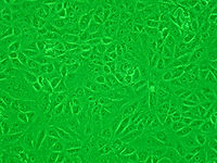
Vero细胞
Vero细胞亦称绿猴肾细胞[1],是一种非整倍性的非洲绿猴(属名:Chlorocebus)肾细胞系,最初由日本千叶大学的安村美博于1962年3月27日,分离自正常成年非洲绿猴的肾脏上皮细胞[2]。“vero”即“verda reno”的缩写,其中“verda reno”在世界语中有“绿色的肾脏”的意思,而“vero”则在世界语中表示“真相”[3] 。

特征
编辑Vero细胞是连续的非整倍性细胞系,这意味着它的染色体数目异常,而且已知连续的细胞系可以通过许多分裂周期,不会老化,Vero细胞的干扰素分泌出现缺陷。它们与正常的哺乳动物细胞不同,在被病毒感染时不会分泌干扰素α/β[4]。然而,它们仍然具有干扰素-α/β受体(IFNAR),因此当将重组干扰素添加到其培养基中时,它们仍然可以作出反应。
Sun等合成了五种不同尺寸的草酸钙(COM)晶体,包括50 nm、200 nm、1 μm、3 μm、10 μm,并且比较这五种尺寸的草酸钙晶体对Vero细胞的损伤差异[5][6]。实验结果表明,COM的尺寸和聚集程度是影响晶体细胞毒性的重要原因,而细胞对晶体的内吞方式与晶体尺寸存在密切的关系[5]。Vero细胞内吞50nm和100 nm的COM晶体主要以网格蛋白介导的途径,内吞1 μm的COM晶体则主要以巨胞饮(macropinocytosis)的形式进行内吞作用,而Vero细胞难以内吞尺寸更大的微米晶体[7]。
2014年,有日本的研究人员确定Vero细胞的整个基因组序列[8]。Vero细胞的12号染色体具有纯合的〜9-Mb缺失,导致基因组中I型干扰素基因簇和细胞周期蛋白依赖性激酶抑制剂CDKN2A和CDKN2B的丢失[8]。尽管非洲绿猴先前被归类为草原猴(Cercopithecus aethiops),但是它们已经被归类为绿猴属(Chlorocebus)[9]。有基因组分析表明,Vero细胞源自雌性绿猴(Chlorocebus sabaeus)[8]。
细胞培养
编辑Vero细胞在传代培养时的生长状况良好,并且发现细胞膜界线清晰和胞浆透明度较好的现象。Vero细胞的形态较为完整,细胞增殖的速度较快。Vero细胞传代后的第三天开始形成单层,传代细胞在第七天形成致密单层,此种致密单层在连续培养第十二天后逐渐老化,细胞在第十六天开始从培养瓶壁上脱落[10]。
Vero细胞在转瓶后,可以在细胞培养二十四小时后出现贴壁,细胞在培养三天后可以达到相对静止期,细胞培养的第五天可以长成单层,细胞培养到第十二天时发现其生长致密,而细胞培养至第十四天时则开始出现老化。转瓶后的细胞生长速度比转瓶前缓慢,然而单层细胞持续的时间比转瓶前更持久。进行支原体检查时未发现有支原体的生长及污染。细胞型分析结果表明Vero 细胞的核型没有发现明显的异常之处,而染色体数目也没有明显变化[10]。
研究用途
编辑Vero细胞可以用于多种研究用途。Vero细胞在建立后不久,就被发现对多种类型的病毒高度敏感,其中包括猿猴空泡病毒40[11]、麻疹病毒[12]、风疹病毒[13]、节足动物携带性病毒[14]及腺病毒[15]等。后来被发现也容易感染细菌毒素,包括白喉毒素[16]、不耐热肠毒素(heat-labile enterotoxins)[17]和志贺氏样毒素[18][19]等。
Vero细胞可以筛选大肠埃希氏菌的毒素。在Vero细胞被建立后,这些毒素亦可以称为“Vero毒素”。由于与痢疾志贺氏菌(Shigella dysenteriae)分离出的志贺氏毒素相似,因此后来被称为志贺氏样毒素(Shiga-like toxin)[8]。
Vero细胞又可以作为锥虫目等真核寄生虫的宿主细胞[8]。此外,Vero细胞被广泛应用于病毒感染分子机制研究、疫苗及重组蛋白的生产[20][21][22],世界卫生组织甚至认可其作为疫苗生产细胞系,建议将其作为流感疫苗生产的替代基质。目前已知Vero细胞可以协助生产狂犬病[23]及水貂犬瘟热等疫苗[24],而用Vero细胞培养的流感疫苗可以更好地介导人体产生对流感的免疫应答[25]。
猪流行性腹泻病毒
编辑Hofmann等通过在培养基中添加胰蛋白酶,证实猪流行性腹泻病毒(PEDV)除了能够在天然宿主的初始靶细胞上增殖外,还可以在Vero细胞中增殖。同时又发现胰蛋白酶对PEDV纤突糖蛋白的切割作用,增强病毒对Vero细胞的感染力[26]。Ye等通过构建稳定表达PEDV ORF3蛋白的Vero细胞,发现ORF3蛋白能够促进PEDV的增殖[27]。然而有研究显示不同的结果,例如Chen等发现orf3基因转译的提前终止,有利于PEDV适应Vero细胞,并且可以提高其在Vero细胞上的复制能力[28];而Sun等对非胰蛋白酶依赖PEDV 85-7的Vero细胞的研究表明,PEDV的复制并没有被orf3基因的突变或转译的提前终止显著影响[29]。另外,Li等构建缺失orf3基因的重组PEDV,发现orf3基因缺失株和携有全长orf3基因的重组病毒,在Vero细胞上的滴度相同,故而推测orf3基因不影响其在Vero细胞上的增殖[30]。
参考文献
编辑- ^ 存档副本. [2021-06-24]. (原始内容存档于2021-06-24).
- ^ Yasumura Y, Kawakita M. The research for the SV40 by means of tissue culture technique. Nippon Rinsho. 1963, 21 (6): 1201–1219 [2020-02-22]. (原始内容存档于2021-02-17).
- ^ Shimizu B. Seno K, Koyama H, Kuroki T , 编. Manual of selected cultured cell lines for bioscience and biotechnology. Tokyo: Kyoritsu Shuppan. 1993: 299–300 [2020-02-22]. ISBN 978-4-320-05386-1. (原始内容存档于2021-02-17) (日语).
- ^ Desmyter, J.; Melnick, J. L.; Rawls, W. E. Defectiveness of interferon production and of rubella virus interference in a line of African green monkey kidney cells (Vero). Journal of Virology. 1968-10, 2 (10): 955–961 [2021-05-09]. ISSN 0022-538X. PMC 375423 . PMID 4302013. doi:10.1128/JVI.2.10.955-961.1968. (原始内容存档于2021-05-25).
- ^ 5.0 5.1 Sun, Xin-Yuan; Gan, Qiong-Zhi; Ouyang, Jian-Ming. Size-dependent cellular uptake mechanism and cytotoxicity toward calcium oxalate on Vero cells. Scientific Reports. 2017-02, 7 (1): 41949 [2021-05-09]. ISSN 2045-2322. PMC 5288769 . PMID 28150811. doi:10.1038/srep41949. (原始内容存档于2017-03-26) (英语).
- ^ Sun, Xin-Yuan; Ouyang, Jian-Ming; Gan, Qiong-Zhi; Liu, Ai-Jie. Renal Epithelial Cell Injury Induced by Calcium Oxalate Monohydrate Depends on their Structural Features: Size, Surface, and Crystalline Structure. Journal of Biomedical Nanotechnology. 2016-11, 12 (11): 2001–2014 [2020-02-22]. ISSN 1550-7033. PMID 29364612. doi:10.1166/jbn.2016.2289. (原始内容存档于2021-05-09).
- ^ 饶晨颖; 郭达; 孙新园; 欧阳健明. 納米和微米一水草酸鈣對Vero細胞毒性的濃度效應. 无机化学学报. 2019, (3): 467-476.
- ^ 8.0 8.1 8.2 8.3 8.4 Osada N, Kohara A, Yamaji T, Hirayama N, Kasai F, Sekizuka T, Kuroda M, Hanada K. The genome landscape of the African green monkey kidney-derived Vero cell line. DNA Research. 2014, 21 (6): 673–83. PMC 4263300 . PMID 25267831. doi:10.1093/dnares/dsu029.
- ^ Haus, Tanja; Akom, Emmanuel; Agwanda, Bernard; Hofreiter, Michael; Roos, Christian; Zinner, Dietmar. Mitochondrial Diversity and Distribution of African Green Monkeys ( Chlorocebus Gray, 1870). American Journal of Primatology. 2013-04, 75 (4): 350–360 [2021-05-09]. ISSN 0275-2565. PMC 3613741 . PMID 23307319. doi:10.1002/ajp.22113. (原始内容存档于2021-05-15) (英语).
- ^ 10.0 10.1 陈静行; 张文杰; 邹超杰. Vero細胞培養特性的研究. 医学美学美容. 2019, 28 (16): 26.
- ^ Yasumura Y; Kawakita Y. Studies on SV40 in tissue culture - preliminary step for cancer research in vitro. Nihon Risnsho 21. 1963: 1201–1215 [2020-02-22].
- ^ Sasaki, K.; Makino, S.; Kasahara, S. Studies on measles virus. II. Propagation in two established simian renal cell lines and development of a plaque assay. The Kitasato Archives of Experimental Medicine. 1964-12, 37 (1): 27–42 [2020-02-22]. ISSN 0023-1924. PMID 5833688. (原始内容存档于2021-05-09).
- ^ Liebhaber, H.; Riordan, J. T.; Horstmann, D. M. Replication of rubella virus in a continuous line of African green monkey kidney cells (Vero). Proceedings of the Society for Experimental Biology and Medicine. Society for Experimental Biology and Medicine (New York, N.Y.). 1967-06, 125 (2): 636–643 [2020-02-22]. ISSN 0037-9727. PMID 4961494. doi:10.3181/00379727-125-32167. (原始内容存档于2020-02-22).
- ^ Simizu, B.; Rhim, J. S.; Wiebenga, N. H. Characterization of the Tacaribe group of arboviruses. I. Propagation and plaque assay of Tacaribe virus in a line of African green monkey kidney cells (Vero). Proceedings of the Society for Experimental Biology and Medicine. Society for Experimental Biology and Medicine (New York, N.Y.). 1967-05, 125 (1): 119–123 [2020-02-22]. ISSN 0037-9727. PMID 6027511. doi:10.3181/00379727-125-32029. (原始内容存档于2021-05-12).
- ^ Rhim, J. S.; Schell, K.; Creasy, B.; Case, W. Biological characteristics and viral susceptibility of an African green monkey kidney cell line (Vero). Proceedings of the Society for Experimental Biology and Medicine. Society for Experimental Biology and Medicine (New York, N.Y.). 1969-11, 132 (2): 670–678 [2020-02-22]. ISSN 0037-9727. PMID 4982209. doi:10.3181/00379727-132-34285. (原始内容存档于2021-05-13).
- ^ Miyamura, K.; Nishio, S.; Ito, A.; Murata, R.; Kono, R. Micro cell culture method for determination of diphtheria toxin and antitoxin titres using VERO cells. I. Studies on factors affecting the toxin and antitoxin titration. Journal of Biological Standardization. 1974-07, 2 (3): 189–201 [2020-02-22]. ISSN 0092-1157. PMID 4214816. doi:10.1016/0092-1157(74)90015-8. (原始内容存档于2020-11-22).
- ^ Speirs, J. I.; Stavric, S.; Konowalchuk, J. Assay of Escherichia coli heat-labile enterotoxin with vero cells. Infection and Immunity. 1977-05, 16 (2): 617–622 [2020-02-22]. ISSN 0019-9567. PMC 421001 . PMID 405326. doi:10.1128/IAI.16.2.617-622.1977. (原始内容存档于2021-05-13).
- ^ Konowalchuk, J.; Speirs, J. I.; Stavric, S. Vero response to a cytotoxin of Escherichia coli. Infection and Immunity. 1977-12, 18 (3): 775–779 [2020-02-22]. ISSN 0019-9567. PMC 421302 . PMID 338490. doi:10.1128/IAI.18.3.775-779.1977. (原始内容存档于2021-05-12).
- ^ Remis, R. S.; MacDonald, K. L.; Riley, L. W.; Puhr, N. D.; Wells, J. G.; Davis, B. R.; Blake, P. A.; Cohen, M. L. Sporadic cases of hemorrhagic colitis associated with Escherichia coli O157:H7. Annals of Internal Medicine. 1984-11, 101 (5): 624–626 [2020-02-22]. ISSN 0003-4819. PMID 6385798. doi:10.7326/0003-4819-101-5-624. (原始内容存档于2020-02-22).
- ^ Nikolay, Alexander; Castilho, Leda R.; Reichl, Udo; Genzel, Yvonne. Propagation of Brazilian Zika virus strains in static and suspension cultures using Vero and BHK cells. Vaccine. 2018-05-24, 36 (22): 3140–3145 [2020-02-22]. ISSN 1873-2518. PMID 28343780. doi:10.1016/j.vaccine.2017.03.018. (原始内容存档于2021-05-09).
- ^ Castrillón-Betancur, Juan Camilo; Urcuqui-Inchima, Silvio. Overexpression of miR-484 and miR-744 in Vero cells alters Dengue virus replication. Memorias Do Instituto Oswaldo Cruz. 2017-04, 112 (4): 281–291 [2020-02-22]. ISSN 1678-8060. PMC 5354610 . PMID 28327787. doi:10.1590/0074-02760160404. (原始内容存档于2021-05-14).
- ^ Kulkarni, Prasad S.; Sahai, Ashish; Gunale, Bhagwat; Dhere, Rajeev M. Development of a new purified vero cell rabies vaccine (Rabivax-S) at the serum institute of India Pvt Ltd. Expert Review of Vaccines. 2017-04, 16 (4): 303–311 [2020-02-22]. ISSN 1744-8395. PMID 28276304. doi:10.1080/14760584.2017.1294068. (原始内容存档于2021-05-09).
- ^ Hassanzadeh, S. Mehdi; Zavareh, Ali; Shokrgozar, M. Ali; Ramezani, Ali; Fayaz, Ahmad. High vero cell density and rabies virus proliferation on fibracel disks versus cytodex-1 in spinner flask. Pakistan journal of biological sciences: PJBS. 2011-04-01, 14 (7): 441–448 [2020-02-22]. ISSN 1028-8880. PMID 21902056. doi:10.3923/pjbs.2011.441.448. (原始内容存档于2020-02-22).
- ^ 冯二凯; 易立; 罗国良; 王振军; 郭利; 陈立志; 程世鹏; 程悦宁. 水貂犬瘟熱Vero細胞活疫苗(CDV3-CL株,懸浮培養)安全性評價. 中国兽药杂志. 2019, 53 (8): 15-22.
- ^ Mochalova, Larisa; Gambaryan, Alexandra; Romanova, Julia; Tuzikov, Alexander; Chinarev, Alexander; Katinger, Dietmar; Katinger, Herman; Egorov, Andrej; Bovin, Nicolai. Receptor-binding properties of modern human influenza viruses primarily isolated in Vero and MDCK cells and chicken embryonated eggs. Virology. 2003-09-01, 313 (2): 473–480 [2020-02-22]. ISSN 0042-6822. PMID 12954214. doi:10.1016/s0042-6822(03)00377-5. (原始内容存档于2021-05-09).
- ^ Hofmann, M.; Wyler, R. Propagation of the virus of porcine epidemic diarrhea in cell culture. Journal of Clinical Microbiology. 1988-11, 26 (11): 2235–2239 [2020-02-22]. ISSN 0095-1137. PMC 266866 . PMID 2853174. doi:10.1128/JCM.26.11.2235-2239.1988. (原始内容存档于2021-05-13).
- ^ Ye, Shiyi; Li, Zhonghua; Chen, Fangzhou; Li, Wentao; Guo, Xiaozhen; Hu, Han; He, Qigai. Porcine epidemic diarrhea virus ORF3 gene prolongs S-phase, facilitates formation of vesicles and promotes the proliferation of attenuated PEDV. Virus Genes. 2015-12, 51 (3): 385–392 [2020-02-22]. ISSN 1572-994X. PMC 7088884 . PMID 26531166. doi:10.1007/s11262-015-1257-y. (原始内容存档于2020-02-22).
- ^ Chen, Fangzhou; Zhu, Yinxing; Wu, Meizhou; Ku, Xugang; Ye, Shiyi; Li, Zhonghua; Guo, Xiaozhen; He, Qigai. Comparative Genomic Analysis of Classical and Variant Virulent Parental/Attenuated Strains of Porcine Epidemic Diarrhea Virus. Viruses. 2015-10-23, 7 (10): 5525–5538 [2021-05-09]. ISSN 1999-4915. PMC 4632399 . PMID 26512689. doi:10.3390/v7102891. (原始内容存档于2020-07-25) (英语).
- ^ Sun, Min; Ma, Jiale; Yu, Zeyanqiu; Pan, Zihao; Lu, Chengping; Yao, Huochun. Identification of two mutation sites in spike and envelope proteins mediating optimal cellular infection of porcine epidemic diarrhea virus from different pathways. Veterinary Research. 2017-12, 48 (1): 44 [2021-05-09]. ISSN 1297-9716. PMC 5577753 . PMID 28854955. doi:10.1186/s13567-017-0449-y. (原始内容存档于2020-08-14) (英语).
- ^ Li, Chunhua; Li, Zhen; Zou, Yong; Wicht, Oliver; van Kuppeveld, Frank J. M.; Rottier, Peter J. M.; Bosch, Berend Jan. Qiu, Jianming , 编. Manipulation of the Porcine Epidemic Diarrhea Virus Genome Using Targeted RNA Recombination. PLoS ONE. 2013-08-02, 8 (8): e69997. ISSN 1932-6203. PMC 3732256 . PMID 23936367. doi:10.1371/journal.pone.0069997 (英语).