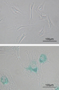
成纤维细胞
成纤维细胞[1][2](fibroblast)又称纤维母细胞,是一种广泛存在于疏松结缔组织或纤维结缔组织内,由中胚层来源的细胞[3],能够分泌胶原和其他细胞外基质以维持结缔组织结构的完整性,在间质更新和创伤修复中发挥重要作用。成纤维细胞的形态多样,有梭形、多突的纺锤形、大多角形和扁平星形等。
| 成纤维细胞 | |
|---|---|
 | |
| 基本信息 | |
| 位置 | 结缔组织 |
| 功能 | 制造细胞外基质和胶原蛋白 |
| 标识字符 | |
| 拉丁文 | fibroblastus |
| MeSH | D005347 |
| TH | H2.00.03.0.01002 |
| FMA | FMA:63877 |
| 《显微解剖学术语》 [在维基数据上编辑] | |

成纤维细胞最初由德国病理学家鲁道夫·菲尔绍与法国解剖学家马蒂亚斯·杜瓦尔于19世纪中叶描述[4]。成纤维细胞与伤口愈合[5]、纤维化[6]等生理学或病理学过程密切相关。组织学中,一般将结缔组织中活跃分泌细胞外基质的细胞称为成纤维细胞,而相对不活跃的纤维细胞则可能是成纤维细胞的前体细胞[7]。
成纤维细胞是一类异质性很强的细胞,且没有明确的表面标记物可用于定义成纤维细胞类群。不同成纤维细胞之间可能发育来源不尽相同,且发挥截然不同的生物学功能[4][8]。
胚胎来源
编辑在胚胎发育中,成纤维细胞和其他的结缔组织一样来源于中胚层。
功能
编辑成纤维细胞的主要功能是分泌细胞外间质的前体,以维持结缔组织结构的完整性。成纤维细胞分泌各类基质和多种纤维。基质的组成决定结缔组织的物理特性。
在形态学上,成纤维细胞具多样性,其形态取决于其所处的位置和活动性。与上皮细胞不同,成纤维细胞不形成细胞单层。但是可以缓慢地迁移。
疾病
编辑成纤维细胞在许多纤维化的疾病中起重要作用,如肺纤维化[9][10]、肾纤维化[11]、和硬皮病[12]。成纤维细胞也在伤口愈合[13],血管新生中起重要作用。
参见
编辑参考文献
编辑- ^ 存档副本. [2023-12-22]. (原始内容存档于2023-12-22).
- ^ 存档副本. [2023-12-22]. (原始内容存档于2023-12-22).
- ^ Murray, Lynne A.; Knight, Darryl A.; Laurent, Geoffrey J. Fibroblasts: 193–200. 2009. doi:10.1016/B978-0-12-374001-4.00015-8.
- ^ 4.0 4.1 Muhl, Lars; Genové, Guillem; Leptidis, Stefanos; Liu, Jianping; He, Liqun. Single-cell analysis uncovers fibroblast heterogeneity and criteria for fibroblast and mural cell identification and discrimination. Nature Communications. 2020, 11 (1). ISSN 2041-1723. doi:10.1038/s41467-020-17740-1.
- ^ Fibroblasts. [16 August 2018]. (原始内容存档于2021-02-04).
- ^ White, Eric S; Mantovani, Alberto R. Inflammation, wound repair, and fibrosis: reassessing the spectrum of tissue injury and resolution. The Journal of Pathology. 2013, 229 (2): 141–144. ISSN 0022-3417. doi:10.1002/path.4126.
- ^ Chong, Sy Giin; Sato, Seidai; Kolb, Martin; Gauldie, Jack. Fibrocytes and fibroblasts—Where are we now. The International Journal of Biochemistry & Cell Biology. 2019, 116: 105595. ISSN 1357-2725. doi:10.1016/j.biocel.2019.105595.
- ^ LeBleu, Valerie S.; Neilson, Eric G. Origin and functional heterogeneity of fibroblasts. The FASEB Journal. 2020, 34 (3): 3519–3536. ISSN 0892-6638. doi:10.1096/fj.201903188R.
- ^ Phan SH. Fibroblast phenotypes in pulmonary fibrosis. Am J Respir Cell Mol Biol. 2003 Sep;29 (3 Suppl):S87-92
- ^ Trovato-Salinaro A, et al., Altered intercellular communication in lung fibroblast cultures from patients with idiopathic pulmonary fibrosis. Respir Res. 2006 Sep 27;7:122
- ^ Strutz F, Muller GA. Renal fibrosis and the origin of the renal fibroblast. Nephrol Dial Transplant. 2006 Dec;21 (12):3368-70
- ^ Pannu J, Trojanowska M. Recent advances in fibroblast signaling and biology in scleroderma. Curr Opin Rheumatol. 2004 Nov;16 (6):739-45
- ^ Darby IA, Hewitson TD. Fibroblast differentiation in wound healing and fibrosis. Int Rev Cytol. 2007;257:143-79