阴部神经
阴部神经(英语:pudendal nerve)为会阴部的主要神经[1]:274。在感觉方面,该神经负责传送男性及女性外阴部、肛门周围,以及会阴的感觉信息;运动信息方面,则支配了男性及女性的尿道括约肌,和外肛门括约肌。一旦损伤则可能导致排粪失禁,这类损伤常见于分娩后遗症,麻醉也可能造成类似的症状。
| 阴部神经 | |
|---|---|
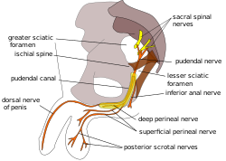
 男性骨盆的阴部神经路径图 | |
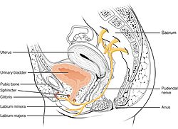
 女性骨盆的阴部神经路径图,阴部神经来自第2、3、4对骶部脊神经(S2、S3、S4),由子宫及肛门之间延伸进入小阴唇、大阴唇及阴蒂 | |
| 基本信息 | |
| 来源 | 荐神经(S2 ~ S4) |
| 走向 | 下直肠神经 会阴神经 阴茎背神经 阴蒂背神经 |
| 标识字符 | |
| 拉丁文 | Nervus pudendus |
| MeSH | D060525 |
| TA98 | A14.2.07.037 |
| TA2 | 6554 |
| FMA | FMA:19037 |
| 格雷氏 | p.967 |
| 《神经解剖学术语》 [在维基数据上编辑] | |
阴部神经会经过闭孔内肌上的阴部管。1836年,爱尔兰解剖学家本杰明·阿尔科克首次描述阴部管,故该管又称阿尔科克氏管(英语:Alcock's canal)。
解剖
编辑阴部神经共有一对,分别在身体的左右侧。每一边的神经都是由三根神经在骶棘韧带及尾骨肌的上边界上方合成一根神经[2]。这三根神经中,中间的及下方的神经合成下方的神经索,因此变成二条神经索,再在骶棘韧带附近联合成一条阴部神经[3]。这三条神经源自第2、3、4对骶部脊神经的腹支,主要是来自第4对骶部脊神经[2][4]:215[5]:157。
阴部神经通过梨状肌及尾骨之间的部位,从坐骨大孔的下方离开骨盆[2]。阴部神经通过骶棘韧带的外侧,从坐骨小孔再进入骨盆,之后会伴随著阴部内动脉及阴部内静脉,沿著坐骨直肠窝的侧壁往上往前,和内动脉和内静脉包覆在闭孔肌筋膜的鞘中,称为阴部管[6]:8。
阴部神经在阴部管内会分支,先分支为内直肠神经,之后是会阴神经,最后是男性的阴茎背神经或是女性的阴蒂的背神经[6]:34。
神经核
编辑此神经是骶丛的主要分支之一[7]:950,神经纤维起源自骶骨段脊髓的欧氏神经核[3]。
变异
编辑阴部神经源自的神经位置也可能会变化,例如有些人的阴部神经可能是源自坐骨神经[8]。,因此坐骨神经的损伤也会影响阴部神经。有时第1节骶神经的后支也可能发展成为阴部神经,甚至是更少见的第5节骶神经(S5)[3]。
功能
编辑阴部神经同时具有运动及感觉两种功能,在自律神经系统方面仅有交感神经的神经纤维,没有副交感神经的纤维[9]:1738。
男性的阴部神经会分出阴茎背神经支配阴茎的感觉,女性则会分出阴蒂背神经支配阴蒂感觉[10]:422。男性的阴囊后侧由阴囊后神经支配其感觉,女性对应的阴唇后神经会支配阴唇的感觉。这些部位有许多神经传导其感觉,阴部神经即为其中之一[11]。阴部神经也传导肛管部位的感觉[6]:8。阴部神经会传导阴茎及阴蒂的感觉,因此也是在阴茎勃起及阴蒂勃起过程中的传入神经[12] :147。阴部神经也负责射精相关的功能[13]。
阴部神经的分支也支配会阴及骨盆底的肌肉,包含球海绵体肌、坐骨海绵体肌[11],以及提肛肌(包括肠骨尾骨肌、耻骨尾骨肌、耻骨直肠肌、女性的耻骨阴道肌或是男性的前列腺提肌)[10]:422[14]、外肛门括约肌(透过下肛门分支)[6]:7、以及男性尿道外括约肌或女性尿道外括约肌[10]:424–425。
阴部神经还透过乙酰胆碱释放控制尿道外括约肌的肌张力。当乙酰胆碱的释放量增加,尿道外括约肌内的骨骼肌纤维会收缩,使尿液留在膀胱,反之则能促进排尿[15]。
临床意义
编辑麻醉
编辑阴部麻醉也称为阴部神经阻断,或鞍神经阻断(saddle nerve block),是产科使用的局部麻醉,可在分娩时麻醉阴部[16]。此麻醉方式会在阴道内壁注射利多卡因,目的是要影响阴部神经[17]。
损伤
编辑阴部神经可能会被压缩或是伸展,造成暂时或是永久的神经病变。若阴部神经拉伸了原来长度的12%,可能会造成不可逆的神经受损[6]:655。若盆腔底急性过度拉伸(例如滞产或是难产)或慢性过度拉伸(因便秘造成排便时的慢性拉伸),可能会让阴部神经出现拉伸造成的神经病变[6]。阴部神经卡压也称为阿尔科克氏管症候群(Alcock canal syndrome),是非常少见的疾病,多半发生在职业的自行车选手身上[18]。像糖尿病及多发性硬化症等系统性疾病也可能透过脱髓鞘病或是其他机制使阴部神经受损[6]:37。骨盆腔的肿瘤(最著名的是大型的骶尾部畸胎瘤)或是去除肿瘤的手术都可能造成神经永久的受损[19]。
若单侧的阴部神经病变可能会造成大便失禁,但也有例外[6]:34。
影像
编辑用一般的断层扫描或是核磁共振成像,很难对阴部神经显像。不过透过断层扫描的引导,可以将针插到邻近阴部神经血管束的部位。坐骨棘在断层扫描时很容易识别,因此会插在此一部位。脊椎针会通过臀肌前进,在坐骨棘上前进几个毫米。之后会注射X光的显影剂,让阴部管内的神经更加清楚,也可以确认针插入的位置是否正确。然后会注射可的松到神经中,进行局部麻醉进行确认,也治疗外阴部的慢性疼痛(女性称为外阴疼痛)、骨盆疼痛和肛门直肠疼痛等[20][21]。
神经传导潜伏期试验
编辑阴部神经的延迟时间可以量化,具体的定义是从在感觉神经给电刺激的时间起,到运动神经有讯号使阴部肌肉收缩的时间,时间太长代表神经受损[22]:46。测试时会有两个固定在手指端的刺激电极及两个量测电极(St Mark电极)[22]:46。
历史
编辑阴部神经的拉丁文为“Nervus pudendus”。“Nervus”一词指的是神经;“Pudenda”一词来自拉丁文,意即外生殖器官,乃源自“pudendum”这个字,意思是“带来羞耻的部份”[23]。阴部管也称为阿尔科克氏管(Alcock's canal),得名自1836年首次纪录此一部位的爱尔兰解剖学家班杰明·阿尔科克。阿尔科克在罗伯特·本特利·托德的《生理暨解剖学百科全书》(The Cyclopædia of Anatomy and Physiology) 一书中,于描述髂动脉群的章节内首次提及阴部神经及阴部管[24]。
其他图片
编辑-
男性骨盆,阴部神经位于图中右方。
-
阴部神经支配构造模式图。
-
男性骨盆的阴部神经路径图。
参见
编辑本条目使用了部分解剖术语。
参考文献
编辑- ^ AMR Agur, AF Dalley, JCB Grant. Grant's atlas of anatomy 13th. Philadelphia: Wolters Kluwer Health/Lippincott Williams & Wilkins. 2013. ISBN 978-1-60831-756-1.
- ^ 2.0 2.1 2.2 Standring S (editor in chief). Gray's Anatomy: The Anatomical Basis of Clinical Practice 39th. Elsevier. 2004. ISBN 978-0-443-06676-4.
- ^ 3.0 3.1 3.2 Shafik, A; el-Sherif, M; Youssef, A; Olfat, ES. Surgical anatomy of the pudendal nerve and its clinical implications. Clinical Anatomy. 1995, 8 (2): 110–5. PMID 7712320. doi:10.1002/ca.980080205.
- ^ Moore, Keith L. Moore, Anne M.R. Agur ; in collaboration with and with content provided by Arthur F. Dalley II ; with the expertise of medical illustrator Valerie Oxorn and the developmental assistance of Marion E. Essential clinical anatomy 3rd. Baltimore, MD: Lippincott Williams & Wilkins. 2007. ISBN 978-0-7817-6274-8.
- ^ Russell RM. Examination of peripheral nerve injuries an anatomical approach. Stuttgart: Thieme. 2006. ISBN 978-3-13-143071-7.
- ^ 6.0 6.1 6.2 6.3 6.4 6.5 6.6 6.7 Wolff BG et al. (编). The ASCRS textbook of colon and rectal surgery. New York: Springer. 2007. ISBN 0-387-24846-3.
- ^ TL King; MC Brucker; JM Kriebs; JO Fahey. Varney's midwifery Fifth. Jones & Bartlett Publishers. 2013. ISBN 978-1-284-02542-2.
- ^ Nayak, Soubhagya R.; Madhan Kumar, S.J.; Krishnamurthy, Ashwin; Latha Prabhu, V.; D'costa, Sujatha; Jetti, Raghu. Unusual origin of dorsal nerve of penis and abnormal formation of pudendal nerve—Clinical significance. Annals of Anatomy - Anatomischer Anzeiger. November 2006, 188 (6): 565–566. doi:10.1016/j.aanat.2006.06.011.
- ^ Neill, editor-in-chief, Jimmy D. Knobil and Neill's physiology of reproduction 3rd. Amsterdam: Elsevier. 2006. ISBN 0-12-515400-3.
- ^ 10.0 10.1 10.2 Drake, Richard L.; Vogl, Wayne; Tibbitts, Adam W.M. Mitchell; illustrations by Richard; Richardson, Paul. Gray's anatomy for students. Philadelphia: Elsevier/Churchill Livingstone. 2005. ISBN 978-0-8089-2306-0.
- ^ 11.0 11.1 Ort, Bruce Ian Bogart, Victoria. Elsevier's integrated anatomy and embryology. Philadelphia, Pa.: Elsevier Saunders. 2007. ISBN 978-1-4160-3165-9.
- ^ Babayan, Mike B. Siroky, Robert D. Oates, Richard K. Handbook of urology diagnosis and therapy 3rd. Philadelphia, PA: Lippincott Williams & Wilkins. 2004. ISBN 978-0-7817-4221-4.
- ^ Penson, David F. Male Sexual Function: A Guide to Clinical Management. Annals of Internal Medicine. 2002.
- ^ Guaderrama, Noelani M.; Liu, Jianmin; Nager, Charles W.; Pretorius, Dolores H.; Sheean, Geoff; Kassab, Ghada; Mittal, Ravinder K. Evidence for the Innervation of Pelvic Floor Muscles by the Pudendal Nerve. Obstetrics & Gynecology. October 2005, 106 (4): 774–781. doi:10.1097/01.AOG.0000175165.46481.a8.
- ^ Fowler, CJ; Griffiths, D; de Groat, WC. The neural control of micturition. Nat. Rev. Neurosci. June 2008, 9: 453–66. PMC 2897743 . PMID 18490916. doi:10.1038/nrn2401.
- ^ Lynna Y. Littleton; Joan Engebretson. Maternal, Neonatal, and Women's Health Nursing, Volume 1. Cengage Learning. 2002: 727.
- ^ Satpathy, Hemant K.; et al. Isaacs, Christine; et al , 编. Transvaginal Pudendal Nerve Block. WebMD LLC. [2015-07-19]. (原始内容存档于2019-07-28).
- ^ Mellion MB. Common cycling injuries. Management and prevention. Sports Med. January 1991, 11 (1): 52–70. PMID 2011683. doi:10.2165/00007256-199111010-00004.
- ^ Lim, Jit F.; Tjandra, Joe J.; Hiscock, Richard; Chao, Michael W. T.; Gibbs, Peter. Preoperative Chemoradiation for Rectal Cancer Causes Prolonged Pudendal Nerve Terminal Motor Latency. Diseases of the Colon & Rectum: 12–19. doi:10.1007/s10350-005-0221-7.
- ^ Calvillo O, Skaribas IM, Rockett C.; Skaribas; Rockett. Computed tomography-guided pudendal nerve block. A new diagnostic approach to long-term anoperineal pain: a report of two cases. Reg Anesth Pain Med. 2000, 25 (4): 420–3. PMID 10925942. doi:10.1053/rapm.2000.7620.
- ^ Hough DM, Wittenberg KH, Pawlina W, Maus TP, King BF, Vrtiska TJ, Farrell MA, Antolak SJ Jr.; Wittenberg; Pawlina; Maus; King; Vrtiska; Farrell; Antolak Jr. Chronic perineal pain caused by pudendal nerve entrapment: anatomy and CT-guided perineural injection technique. Am J Roentgenol. 2003, 181 (2): 561–7. PMID 12876048. doi:10.2214/ajr.181.2.1810561.
- ^ 22.0 22.1 G.A. Santoro, A.P. Wieczorek, C.I. Bartram (editors). Pelvic floor disorders imaging and multidisciplinary approach to management. Dordrecht: Springer. 2010. ISBN 978-88-470-1542-5.
- ^ Harper, Douglas. Pudendum. Online Etymology Dictionary. [2014-02-28]. (原始内容存档于2017-01-18).
- ^ Oelhafen, Kim; Shayota, Brian J.; Muhleman, Mitchel; Klaassen, Zachary; Tubbs, R. Shane; Loukas, Marios. Benjamin Alcock (1801-?) and his canal. Clinical Anatomy. 2013-09, 26 (6): 662–666. PMID 22488487. doi:10.1002/ca.22080.
外部链接
编辑- Anatomy figure: 41:04-11 at Human Anatomy Online, SUNY Downstate Medical Center - 女性会阴下视观,以及内阴动脉的分支
- figures/chapter_32/32-2.HTM — Basic Human Anatomy at Dartmouth Medical School
- figures/chapter_32/32-3.HTM — Basic Human Anatomy at Dartmouth Medical School
- 横截面图像:pelvis/pelvis-female-17 - 维也纳医科大学生物塑化实验室提供
- www.nervemed.com 上的诊断与治疗方法
- www.pudendal.com (页面存档备份,存于互联网档案馆)
- 阴部神经压迫症候群 - chronicprostatitis.com (页面存档备份,存于互联网档案馆)
- 阴部神经阻断的电脑断层影像