T细胞
T细胞(英语:T cell;T lymphocyte)是淋巴细胞的一种,在免疫反应中扮演着重要的角色,T是胸腺(thymus)的英文缩写。T细胞在骨髓被制造出来之后,在胸腺内进行“新兵训练”分化成熟为不同亚型的效应T细胞,成熟后就移居于周围淋巴组织中开始工作。
| T细胞 | |
|---|---|
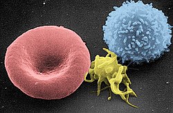
 人类T细胞的扫描电子显微镜图 | |
 | |
| 基本信息 | |
| 系统 | 免疫系统 |
| 标识字符 | |
| 拉丁文 | lymphocytus T |
| MeSH | D013601 |
| TH | H2.00.04.1.02007 |
| FMA | FMA:62870 |
| 《显微解剖学术语》 [在维基数据上编辑] | |
T细胞膜表面分子有两大类:T细胞受体(TCR)和分化群分子(CD molecules),其与T细胞的功能相关,也是T细胞的表面标志(cell-surface marker),可以用以分离、鉴定不同亚群的T细胞[1]。
发育
编辑起源
编辑所有的T细胞都来源于造血干细胞(HSC),造血干细胞会分化为多能祖细胞(MPP),多能祖细胞又会分化为共同淋巴祖细胞(CLP),之后CLP只有三种分化路径,即T细胞、B细胞和NK细胞[2]。 那些分化为T细胞的CLP将会随着血流到达胸腺,并成为早期胸腺祖细胞(ETP),现在这些细胞既不表达CD4也不表达CD8[3]。这些细胞将经过一轮分裂之后会进入DN1阶段。
TCR-β选择
编辑在DN2阶段(CD44+CD25+),细胞上调RAG1/2并重排TCR(T细胞受体)-β基因座,V-D-J序列和恒定区序列,目的是产生一个有功能的TCR-β链。当细胞经过DN3阶段(CD44-CD25+)时,细胞将会和TCRβ一起表达一个未经重排的α-链(pre-Tα),如果重排后的β-链可以和pre-Tα形成二聚体,细胞将产生信号停止β-链的重排[4]。虽然这个信号需要pre-TCR在细胞膜上表达,不过它与pre-TCR和配体的结合无关。如果pre-TCR形成了,细胞会下调CD25并进入DN4阶段(CD25-CD44-),这些细胞将继续分裂并重排TCRα的基因座。
阳性选择
编辑双阳性(CD4+/CD8+)的T细胞会向胸腺皮层深处迁移,并会接触到胸腺皮层上皮细胞表面的“自体抗原”(self-antigens)。这些自体抗原结合在胸腺上皮细胞表面的MHC分子上,只有与胸腺细胞的MHC分子表现出足够强的结合力的T细胞,才能接收到必要的“存活信号”,而无法接收到足够“存活信号”的T细胞将会凋亡。在这个持续几天的阳性选择过程中,大部分的T细胞都会死去[5]。
一个T细胞的命运就在阳性选择的过程中被决定。在双阳性(CD4+/CD8+)T细胞中,能够与MHC Ⅱ类分子结合得较好的将成为CD4+细胞,而和MHC Ⅰ类分子有更高亲和力的将成为CD8+细胞。将成为CD4+细胞的细胞将会逐渐下调自己的CD8,最终成为单阳性的CD4+细胞[6]。
阴性选择
编辑在阳性选择中存活下来的T细胞将会向胸腺皮质边缘和髓质区迁移;在髓质区,它们又会接触到胸腺髓质上皮细胞(mTECs)表面的自体抗原[7]。mTECs会在它们的MHC Ⅰ类分子上呈递来自全身各个组织的自体抗原。一些mTECs被胸腺树状细胞吞噬,它们的自体抗原就会呈递在树状细胞的MHC Ⅱ类分子上(经过了阳性选择的CD4+细胞只能识别MHC Ⅱ类分子)。在这里,与自体抗原表现出过强的亲和力的T细胞会接收到凋亡信号并凋亡(在这些细胞中也有一部分会成为调节T细胞),存活下来的细胞就作为成熟的初级T细胞离开胸腺[8]。这一过程是中枢免疫耐受的重要组成部分,其意义在于筛选掉可能对自体抗原产生反应的T细胞,从而避免自体免疫疾病的发生。
胸腺输出
编辑经过阳性选择和阴性选择,最初到达胸腺的T细胞中有98%死亡,存活下来的2%成为了具有成熟免疫功能的T细胞。胸腺产生成熟T细胞的数量大致随着个体衰老而减少,在中年人的体内,胸腺的大小平均每年缩小3%[9]。所以,对中老年人而言,外周T细胞的增殖和再生对于免疫系统的意义更大。
分类
编辑T细胞根据功能的差异被分为几个亚型。虽然在胸腺中就分化出了CD4+和CD8+两者,但是在外周T细胞还会发生进一步的分化。
常规适应性T细胞
编辑辅助性CD4+ T细胞
编辑辅助性T细胞(TH细胞)对其他淋巴细胞的活动起辅助作用,包括B细胞向浆细胞和记忆B细胞的发育,以及细胞毒性T细胞和巨噬细胞的激活。它们也被称为CD4+ T细胞,因为它们的细胞表面有CD4蛋白的表达。辅助T细胞在遇到抗原呈递细胞(APC)表面MHC-II分子结合的外部抗原时被激活,一旦被激活就会快速分裂并开始分泌调节免疫反应的细胞因子。辅助T细胞在受到不同细胞因子刺激的情况下,也会进一步分化成不同亚型的辅助T细胞[10]。它们是已知的HIV病毒的目标细胞,在艾滋病发病时会急剧减少。滤泡辅助性T细胞是参与体液应答的CD4阳性辅助性T细胞的一个亚群[11],属一类专门为B细胞提供帮助的CD4+ T细胞[12]。
调节性CD4+ T细胞
编辑调节性T细胞(Treg细胞)旧称抑制性T细胞,对于免疫耐受至关重要。它们的主要工作就是及时有效的结束免疫反应,以及抑制那些从阴性筛选中逃逸的自体免疫T细胞,防止免疫反应对抑制性机体自身造成过度损害。
调节性T细胞既可以在胸腺中发育分化完成,称为胸腺调节T细胞;也可以在外周组织受免疫反应诱导分化,称为外周调节T细胞[13]。两者都表达FOXP3作为其细胞表面标志物,FOXP3基因的突变会影响调节T细胞的发育,并诱发致命的自体免疫病IPEX。
其他几类不表达FOXP3基因的T细胞具有免疫抑制作用,例如Tr1细胞和Th3细胞。Tr1与IL-10相关,Th3与TGF-beta相关。最近,Th17细胞也被列入此类免疫抑制细胞之中[14]。
细胞毒性CD8+ T细胞
编辑细胞毒性T细胞(CTLs, killer T cells)负责杀伤被病毒感染的细胞和癌细胞,在对器官移植的免疫排斥中也有参与。其特点在于细胞表面的CD8蛋白质。它通过识别所有有核细胞表面的MHC-I分子上的短肽抗原,来分辨正常细胞和应杀伤的异常细胞。细胞毒性T细胞还可分泌重要的细胞因子IL-2和IFNγ,来影响其他免疫细胞的功能,特别是巨噬细胞和NK细胞。
记忆性T细胞
编辑还未结合过外部抗原的初级T细胞,一旦结合了抗原呈递细胞表面MHC分子所包裹的外部抗原,就会开始增殖分化为效应T细胞和记忆T细胞(其他信号适当的共刺激对这一过程也是必要的)。曾经,人们认为记忆T细胞只分为中央记忆T细胞和效应记忆T细胞[15]。但是之后,新的记忆T细胞种类不断被发现,例如组织驻留记忆T细胞 (Trm)等等。记忆T细胞的共同特点在于其寿命较长(可长达数十年),而且在识别到特定抗原时可以快速分裂为大量的效应T细胞。通过这样的方式,记忆T细胞就为人体的免疫系统保存了对之前感染过病原体的“记忆”。记忆T既可以是CD4+也可以是CD8+,一般会表达CD45RO[16]。
类固有T细胞
编辑自然杀伤T细胞
编辑自然杀伤T细胞(NKT细胞),请不要把它和固有免疫中的自然杀伤细胞(NK细胞)混淆。 与一般T细胞识别MHC分子上的肽链抗原不同,NKT识别的是CD1d分子上结合的糖蛋白抗原。被激活后,它们可以执行类似辅助T细胞和细胞毒性T细胞的功能,即释放细胞因子和细胞毒素。有证据表明,它们能够识别并杀灭某些肿瘤细胞和被疱疹病毒感染的细胞[17]。
粘膜相关的不变T细胞
编辑粘膜相关的不变T细胞(MAIT)具有固有免疫效应细胞的特质[18][19]。在人体内,MAIT细胞分布于血液、肝脏、肺部、黏膜,具有抵御微生物感染的能力[18]。MHC-I的类似物,MR1,可以向MAIT细胞呈递细菌产生的代谢物抗原[20][21][22]。接受了MR1呈递的外部抗原后,MAIT细胞可以释放促炎症细胞因子并裂解被细菌感染的细胞[18][22]。MAIT也可以通过不依靠MR1的信号通路激活[22]。除了表现出类似固有免疫的功能外,MAIT细胞也辅助获得性免疫反应,甚至表现出类似记忆细胞的特征[18]。此外,MAIT也被认为在自体免疫病中发挥作用,如多发性硬化、风湿和炎症性肠病[23][24],虽然决定性的证据还有待发现[25][26][27][28]。
γδ T细胞
编辑γδ T细胞代表了T细胞中一小部分不表达αβ-TCR而表达γδ-TCR的类型,在小鼠和人体内仅占全部T细胞的2%;在兔子、绵羊和鸡体内,γδ T细胞占全部T细胞的比例则可能高达60%。它们主要分布在肠道粘膜,作为一类上皮内淋巴细胞。关于其抗原识别的细节我们仍知之甚少,不过似乎γδ T细胞并不受限于MHC分子的呈递。特别的是,γδ T细胞能够对一类磷酸类抗原做出快速的反应,而这类抗原物质在各种细胞(细菌、植物、癌细胞等)中都有发现。
活化
编辑CD4+ T细胞的激活需要T细胞上的TCR和共受体(CD28或ICOS),抗原呈递细胞上的MHCII和共激活分子两对分子的分别,同时结合。仅其中一对的结合,无法产生有效的T细胞激活。理想的CD8+ T细胞激活则依赖于CD4+ T细胞的信号转导[30]。CD4+细胞可以在初级CD8 T细胞的初次免疫应答中给予帮助,并且在急性感染的后期维持CD8+ 记忆T细胞的活性。所以,CD4+ T的激活对于CD8+ T细胞的活动是有利的[31][32][33]。
相比于MHC分子上的抗原,抗原呈递细胞的共激活分子一般是由病原体的副产物、热休克蛋白或者坏死的细胞碎片诱导表达的。共刺激机制被认为可以避免自体免疫的发生,因为即使T细胞错误地结合了自体抗原,也可能因为没有受到合适的共刺激而无法正常活化。一旦T细胞被正确地活化,它的细胞表面蛋白表达就会发生巨大的改变,活化T细胞的标志蛋白包括CD69,CD71,CD25 (也是调节T细胞的标志)和HLA-DR (人类T细胞的特异标志)。CTLA-4在活化T细胞表面的上调,对共激活受体有竞争性抑制作用,可以避免活化T细胞的过度活化。活化T细胞的表面糖基化情况也有改变[34]。
T细胞受体(TCR)是由几种蛋白质组合成的复合体。TCR的两个主要组分是由两个独立基因分别编码的TCRα和TCRβ,其他的组分包括CD3家族的蛋白:CD3εγ和CD3εδ的异二聚体,以及最重要的CD3ζ同二聚体。CD3ζ同二聚体上共有6个ITAM基序,可被磷酸化并启动一系列级联反应,导致TCR复合体的聚集。
虽然在绝大部分情况下T细胞活化都依赖于TCR对抗原的识别,其他的活化途径也有被发现,例如细胞毒性T细胞可以被其他CD8 T细胞识别并导致自身的极化[35]。T细胞活化的过程也受到活性氧类物质的影响[36]。
抗原识别
编辑T细胞的主要特点就是能够分辨正常细胞和异常细胞的能力[37]。不论是正常细胞还是异常细胞,都会表达大量的MHC-抗原多肽复合体(pMHC)。虽然T细胞与正常细胞的pMHC有一定结合力,但是T细胞并不会被激活;但即使异常细胞的pMHC与正常细胞只有细微的差别,也能够刺激T细胞发生免疫反应。这样对不同抗原完全不同的反应特征称为T细胞的抗原识别,关于这一机理实现的具体细节如今仍然没有定论[38]。
临床意义
编辑缺陷
编辑T细胞缺陷可能意味着T细胞数量的减少或者T细胞功能的缺失。完全的T细胞缺陷可能来自于一些遗传因素,例如严重复合型免疫缺乏症(SCID)、欧门氏症候群或软骨毛发发育不全。[46]部分的T细胞缺陷可能是由于获得性免疫缺陷综合征(AIDS)、遗传性的迪乔治综合征(DGS)、染色体断裂综合征(CBSs),或者B细胞和T细胞的复合缺陷,例如毛细血管扩张性运动失调 (AT) 和欧德里综合征(Wiskott–Aldrich syndrome)[39]。
T细胞缺陷患者面临的主要风险,主要是一些细胞内病原体,例如单纯疱疹病毒、分枝杆菌和李斯特菌。同时,真菌感染在T细胞缺陷患者身上往往也很常见且严重[40]。
癌症
编辑T细胞癌变诱发的肿瘤称为T细胞淋巴瘤,在非霍奇金淋巴瘤中约占10%[41]。
耗竭
编辑T细胞耗竭是一种T细胞功能失常的状态,其表现为进展性的功能丧失、基因表达谱的变化、和抑制性细胞因子的持续分泌。T细胞耗竭可能发生于慢性感染、败血症、癌症的进程中[42]。耗竭的T细胞即使再次暴露于抗原刺激之中也无法恢复正常功能[43]。
慢性感染和败血症
编辑T细胞耗竭的直接原因包括持续的抗原刺激、以及CD4细胞的缺失[44]。长时间的抗原暴露和高病毒负载可以加重T细胞耗竭的程度。2-4周的持续抗原暴露就可导致T细胞耗竭[45]。另一个可以导致T细胞耗竭的因素是包括PD-1在内的一系列抑制性受体[46][47]。细胞因子IL-10或TGF-β也可以导致耗竭[48][49]。调节T细胞因为可以分泌IL-10和TGF-β,也与T细胞耗竭相关[50]。在阻断PD-1受体并减少调节T细胞数量后,T细胞耗竭的情况可以得到反转[51]。[58]在败血症中,抑制性的细胞因子风暴也会造成T细胞耗竭[52][53]。现在已有致力于通过阻断抑制性受体的方式来治疗败血症的疗法研究[54][55][56]。
器官移植
编辑与感染时的情况类似,器官移植带来的持续异种抗原暴露也会造成T细胞耗竭[57]。肾移植后,T细胞应答能力会随时间减弱[58]。这些数据说明T细胞耗竭导致的CD8+ T细胞数量减少可能是器官移植耐受中的重要一环[59]。已有几项研究证明了慢性感染对器官移植后的免疫耐受和长期生存有利,而T细胞耗竭起着一定介导的作用[60][61][62]。虽然已有T细胞耗竭对器官移植有利的证据,但是T细胞耗竭同时带来的感染和癌变风险依然不能忽视[63]。
癌症
编辑在癌症进程中,T细胞耗竭显然对癌组织的存活有利。已有研究证明癌细胞和一些癌症相关细胞可以主动地诱导T细胞耗竭的发生[64][65] [66]。在白血病中,T细胞耗竭也与其复发相关[67]。一些研究甚至提出可以基于T细胞抑制性受体PD-1的表达状态来预测白血病复发的情况[68]。由于免疫抑制性受体与T细胞耗竭以及癌症之间的关系,近年来有大量的研究和临床试验致力于通过阻断免疫抑制性受体来治疗癌症,其中有一些已经被认定有效并投入临床使用[69][70]。
参见
编辑参考文献
编辑- ^ Janeway, Charles. Immunobiology: the immune system in health and disease 5th. New York: Garland Pub. 2001 [2020-02-12]. ISBN 978-0-8153-3642-6. OCLC 45708106. (原始内容存档于2019-10-17).
- ^ Kondo, Motonari. One Niche to Rule Both Maintenance and Loss of Stemness in HSCs. Immunity. 2016-12-20, 45 (6): 1177–1179 [2020-02-23]. ISSN 1097-4180. PMID 28002722. doi:10.1016/j.immuni.2016.12.003. (原始内容存档于2020-03-18).
- ^ Osborne, Lisa C.; Dhanji, Salim; Snow, Jonathan W.; Priatel, John J.; Ma, Melissa C.; Miners, M. Jill; Teh, Hung-Sia; Goldsmith, Mark A.; Abraham, Ninan. Impaired CD8 T cell memory and CD4 T cell primary responses in IL-7R alpha mutant mice. The Journal of Experimental Medicine. 2007-03-19, 204 (3): 619–631 [2020-02-23]. ISSN 0022-1007. PMC 2137912 . PMID 17325202. doi:10.1084/jem.20061871. (原始内容存档于2020-03-18).
- ^ Murphy, Kenneth (Kenneth M.); Walport, Mark.; Janeway, Charles. Janeway's immunobiology 8th. New York: Garland Science. 2012: 301–305. ISBN 978-0-8153-4243-4. OCLC 733935898.
- ^ Starr, Timothy K.; Jameson, Stephen C.; Hogquist, Kristin A. Positive and negative selection of T cells. Annual Review of Immunology. 2003, 21: 139–176 [2020-02-23]. ISSN 0732-0582. PMID 12414722. doi:10.1146/annurev.immunol.21.120601.141107. (原始内容存档于2020-03-18).
- ^ Zerrahn, J.; Held, W.; Raulet, D. H. The MHC reactivity of the T cell repertoire prior to positive and negative selection. Cell. 1997-03-07, 88 (5): 627–636 [2020-02-23]. ISSN 0092-8674. PMID 9054502. doi:10.1016/s0092-8674(00)81905-4. (原始内容存档于2020-03-18).
- ^ Hinterberger, Maria; Aichinger, Martin; Prazeres da Costa, Olivia; Voehringer, David; Hoffmann, Reinhard; Klein, Ludger. Autonomous role of medullary thymic epithelial cells in central CD4(+) T cell tolerance. Nature Immunology. 2010-06, 11 (6): 512–519 [2020-02-23]. ISSN 1529-2916. PMID 20431619. doi:10.1038/ni.1874. (原始内容存档于2020-03-18).
- ^ Pekalski, Marcin L.; García, Arcadio Rubio; Ferreira, Ricardo C.; Rainbow, Daniel B.; Smyth, Deborah J.; Mashar, Meghavi; Brady, Jane; Savinykh, Natalia; Dopico, Xaquin Castro. Neonatal and adult recent thymic emigrants produce IL-8 and express complement receptors CR1 and CR2. JCI insight. 2017-08-17, 2 (16) [2020-02-23]. ISSN 2379-3708. PMC 5621870 . PMID 28814669. doi:10.1172/jci.insight.93739. (原始内容存档于2020-03-18).
- ^ Haynes, B. F.; Markert, M. L.; Sempowski, G. D.; Patel, D. D.; Hale, L. P. The role of the thymus in immune reconstitution in aging, bone marrow transplantation, and HIV-1 infection. Annual Review of Immunology. 2000, 18: 529–560 [2020-02-23]. ISSN 0732-0582. PMID 10837068. doi:10.1146/annurev.immunol.18.1.529. (原始内容存档于2020-03-18).
- ^ Gutcher, Ilona; Becher, Burkhard. APC-derived cytokines and T cell polarization in autoimmune inflammation. The Journal of Clinical Investigation. 2007-05, 117 (5): 1119–1127 [2020-02-23]. ISSN 0021-9738. PMC 1857272 . PMID 17476341. doi:10.1172/JCI31720. (原始内容存档于2020-03-18).
- ^ Crotty S (2014) T follicular helper cell differentiation, function, and roles in disease.Immunity 41:529–542.
- ^ 何岚; 孙兵. 滤泡辅助性T细胞分化和功能的研究进展 (PDF). 生命科学. 2016, 28 (2): 146 [2023-05-05]. doi:10.13376/j.cbls/2016021. (原始内容存档 (PDF)于2023-05-05).
- ^ Abbas, Abul K.; Benoist, Christophe; Bluestone, Jeffrey A.; Campbell, Daniel J.; Ghosh, Sankar; Hori, Shohei; Jiang, Shuiping; Kuchroo, Vijay K.; Mathis, Diane. Regulatory T cells: recommendations to simplify the nomenclature. Nature Immunology. 2013-04, 14 (4): 307–308 [2020-02-23]. ISSN 1529-2916. PMID 23507634. doi:10.1038/ni.2554. (原始内容存档于2020-03-18).
- ^ Singh, Bhagirath; Schwartz, Jordan Ari; Sandrock, Christian; Bellemore, Stacey M.; Nikoopour, Enayat. Modulation of autoimmune diseases by interleukin (IL)-17 producing regulatory T helper (Th17) cells. The Indian Journal of Medical Research. 2013-11, 138 (5): 591–594 [2020-02-23]. ISSN 0971-5916. PMC 3928692 . PMID 24434314. (原始内容存档于2020-03-18).
- ^ Sallusto, F.; Lenig, D.; Förster, R.; Lipp, M.; Lanzavecchia, A. Two subsets of memory T lymphocytes with distinct homing potentials and effector functions. Nature. 1999-10-14, 401 (6754): 708–712 [2020-02-23]. ISSN 0028-0836. PMID 10537110. doi:10.1038/44385. (原始内容存档于2020-03-18).
- ^ Akbar, A. N.; Terry, L.; Timms, A.; Beverley, P. C.; Janossy, G. Loss of CD45R and gain of UCHL1 reactivity is a feature of primed T cells. Journal of Immunology (Baltimore, Md.: 1950). 1988-04-01, 140 (7): 2171–2178 [2020-02-23]. ISSN 0022-1767. PMID 2965180. (原始内容存档于2020-03-18).
- ^ Mallevaey, Thierry; Fontaine, Josette; Breuilh, Laetitia; Paget, Christophe; Castro-Keller, Alexandre; Vendeville, Catherine; Capron, Monique; Leite-de-Moraes, Maria; Trottein, François. Invariant and noninvariant natural killer T cells exert opposite regulatory functions on the immune response during murine schistosomiasis. Infection and Immunity. 2007-05, 75 (5): 2171–2180 [2020-02-23]. ISSN 0019-9567. PMC 1865739 . PMID 17353286. doi:10.1128/IAI.01178-06. (原始内容存档于2020-03-18).
- ^ 18.0 18.1 18.2 18.3 Napier RJ, Adams EJ, Gold MC, Lewinsohn DM. The Role of Mucosal Associated Invariant T Cells in Antimicrobial Immunity. Frontiers in Immunology. 2015-07-06, 6: 344. PMC 4492155 . PMID 26217338. doi:10.3389/fimmu.2015.00344.
- ^ Gold MC, Lewinsohn DM. Mucosal associated invariant T cells and the immune response to infection. Microbes and Infection. August 2011, 13 (8–9): 742–8. PMC 3130845 . PMID 21458588. doi:10.1016/j.micinf.2011.03.007.
- ^ Eckle SB, Corbett AJ, Keller AN, Chen Z, Godfrey DI, Liu L, Mak JY, Fairlie DP, Rossjohn J, McCluskey J. Recognition of Vitamin B Precursors and Byproducts by Mucosal Associated Invariant T Cells. The Journal of Biological Chemistry. December 2015, 290 (51): 30204–11. PMC 4683245 . PMID 26468291. doi:10.1074/jbc.R115.685990.
- ^ Ussher JE, Klenerman P, Willberg CB. Mucosal-associated invariant T-cells: new players in anti-bacterial immunity. Frontiers in Immunology. 2014-10-08, 5: 450. PMC 4189401 . PMID 25339949. doi:10.3389/fimmu.2014.00450.
- ^ 22.0 22.1 22.2 Howson LJ, Salio M, Cerundolo V. MR1-Restricted Mucosal-Associated Invariant T Cells and Their Activation during Infectious Diseases. Frontiers in Immunology. 2015-06-16, 6: 303. PMC 4468870 . PMID 26136743. doi:10.3389/fimmu.2015.00303.
- ^ Hinks TS. Mucosal-associated invariant T cells in autoimmunity, immune-mediated diseases and airways disease. Immunology. May 2016, 148 (1): 1–12. PMC 4819138 . PMID 26778581. doi:10.1111/imm.12582.
- ^ Bianchini E, De Biasi S, Simone AM, Ferraro D, Sola P, Cossarizza A, Pinti M. Invariant natural killer T cells and mucosal-associated invariant T cells in multiple sclerosis. Immunology Letters. March 2017, 183: 1–7. PMID 28119072. doi:10.1016/j.imlet.2017.01.009.
- ^ Serriari NE, Eoche M, Lamotte L, Lion J, Fumery M, Marcelo P, Chatelain D, Barre A, Nguyen-Khac E, Lantz O, Dupas JL, Treiner E. Innate mucosal-associated invariant T (MAIT) cells are activated in inflammatory bowel diseases. Clinical and Experimental Immunology. May 2014, 176 (2): 266–74. PMC 3992039 . PMID 24450998. doi:10.1111/cei.12277.
- ^ Huang S, Martin E, Kim S, Yu L, Soudais C, Fremont DH, Lantz O, Hansen TH. MR1 antigen presentation to mucosal-associated invariant T cells was highly conserved in evolution. Proceedings of the National Academy of Sciences of the United States of America. May 2009, 106 (20): 8290–5. Bibcode:2009PNAS..106.8290H. PMC 2688861 . PMID 19416870. doi:10.1073/pnas.0903196106.
- ^ Chua WJ, Hansen TH. Bacteria, mucosal-associated invariant T cells and MR1. Immunology and Cell Biology. November 2010, 88 (8): 767–9. PMID 20733595. doi:10.1038/icb.2010.104.
- ^ Kjer-Nielsen L, Patel O, Corbett AJ, Le Nours J, Meehan B, Liu L, Bhati M, Chen Z, Kostenko L, Reantragoon R, Williamson NA, Purcell AW, Dudek NL, McConville MJ, O'Hair RA, Khairallah GN, Godfrey DI, Fairlie DP, Rossjohn J, McCluskey J. MR1 presents microbial vitamin B metabolites to MAIT cells (PDF). Nature. November 2012, 491 (7426): 717–23. Bibcode:2012Natur.491..717K. PMID 23051753. doi:10.1038/nature11605.
- ^ The NIAID resource booklet "Understanding the Immune System (pdf)" (页面存档备份,存于互联网档案馆).
- ^ Williams, Matthew A.; Bevan, Michael J. Effector and memory CTL differentiation. Annual Review of Immunology. 2007, 25: 171–192 [2020-02-23]. ISSN 0732-0582. PMID 17129182. doi:10.1146/annurev.immunol.25.022106.141548. (原始内容存档于2020-03-18).
- ^ Janssen EM, Lemmens EE, Wolfe T, Christen U, von Herrath MG, Schoenberger SP. CD4+ T cells are required for secondary expansion and memory in CD8+ T lymphocytes. Nature. February 2003, 421 (6925): 852–6. Bibcode:2003Natur.421..852J. PMID 12594515. doi:10.1038/nature01441.
- ^ Shedlock DJ, Shen H. Requirement for CD4 T cell help in generating functional CD8 T cell memory. Science. April 2003, 300 (5617): 337–9. Bibcode:2003Sci...300..337S. PMID 12690201. doi:10.1126/science.1082305.
- ^ Sun JC, Williams MA, Bevan MJ. CD4+ T cells are required for the maintenance, not programming, of memory CD8+ T cells after acute infection. Nature Immunology. September 2004, 5 (9): 927–33. PMC 2776074 . PMID 15300249. doi:10.1038/ni1105.
- ^ Maverakis E, Kim K, Shimoda M, Gershwin M, Patel F, Wilken R, Raychaudhuri S, Ruhaak LR, Lebrilla CB. Glycans in the immune system and The Altered Glycan Theory of Autoimmunity. J Autoimmun. 2015, 57 (6): 1–13. PMC 4340844 . PMID 25578468. doi:10.1016/j.jaut.2014.12.002.
- ^ Milstein O, Hagin D, Lask A, Reich-Zeliger S, Shezen E, Ophir E, Eidelstein Y, Afik R, Antebi YE, Dustin ML, Reisner Y. CTLs respond with activation and granule secretion when serving as targets for T cell recognition. Blood. January 2011, 117 (3): 1042–52. PMC 3035066 . PMID 21045195. doi:10.1182/blood-2010-05-283770.
- ^ Belikov AV, Schraven B, Simeoni L. T cells and reactive oxygen species. Journal of Biomedical Science. October 2015, 22: 85. PMC 4608155 . PMID 26471060. doi:10.1186/s12929-015-0194-3.
- ^ Feinerman O, Germain RN, Altan-Bonnet G. Quantitative challenges in understanding ligand discrimination by alphabeta T cells. Mol. Immunol. 2008, 45 (3): 619–31. PMC 2131735 . PMID 17825415. doi:10.1016/j.molimm.2007.03.028.
- ^ Dushek O, van der Merwe PA. An induced rebinding model of antigen discrimination. Trends Immunol. 2014, 35 (4): 153–8. PMC 3989030 . PMID 24636916. doi:10.1016/j.it.2014.02.002.
- ^ Medscape > T-cell Disorders (页面存档备份,存于互联网档案馆). Author: Robert A Schwartz, MD, MPH; Chief Editor: Harumi Jyonouchi, MD. Updated: May 16, 2011
- ^ Bannister, Barbara A.; Jones, Jane. Infection : microbiology and management 3rd. Malden, Mass.: Blackwell Pub. 2006: 435. ISBN 978-1-4443-2393-1. OCLC 592756309.
- ^ The Lymphomas (PDF). The Leukemia & Lymphoma Society: 2. May 2006 [2008-04-07]. (原始内容 (PDF)存档于2008-07-06).
- ^ Yi, John S.; Cox, Maureen A.; Zajac, Allan J. T-cell exhaustion: characteristics, causes and conversion. Immunology. 2010-04, 129 (4): 474–481 [2020-02-23]. ISSN 1365-2567. PMC 2842494 . PMID 20201977. doi:10.1111/j.1365-2567.2010.03255.x. (原始内容存档于2020-03-26).
- ^ Wang, Qin; Pan, Wen; Liu, Yanan; Luo, Jinzhuo; Zhu, Dan; Lu, Yinping; Feng, Xuemei; Yang, Xuecheng; Dittmer, Ulf. Hepatitis B Virus-Specific CD8+ T Cells Maintain Functional Exhaustion after Antigen Reexposure in an Acute Activation Immune Environment. Frontiers in Immunology. 2018, 9: 219 [2020-02-23]. ISSN 1664-3224. PMC 5816053 . PMID 29483916. doi:10.3389/fimmu.2018.00219. (原始内容存档于2020-03-26).
- ^ Matloubian, M.; Concepcion, R. J.; Ahmed, R. CD4+ T cells are required to sustain CD8+ cytotoxic T-cell responses during chronic viral infection. Journal of Virology. 1994-12, 68 (12): 8056–8063 [2020-02-23]. ISSN 0022-538X. PMC 237269 . PMID 7966595. (原始内容存档于2020-03-18).
- ^ Angelosanto, Jill M.; Blackburn, Shawn D.; Crawford, Alison; Wherry, E. John. Progressive loss of memory T cell potential and commitment to exhaustion during chronic viral infection. Journal of Virology. 2012-08, 86 (15): 8161–8170 [2020-02-23]. ISSN 1098-5514. PMC 3421680 . PMID 22623779. doi:10.1128/JVI.00889-12. (原始内容存档于2020-03-18).
- ^ Wherry EJ. T cell exhaustion. Nature Immunology. June 2011, 12 (6): 492–9. PMID 21739672. doi:10.1038/ni.2035.
- ^ Okagawa T, Konnai S, Nishimori A, Maekawa N, Goto S, Ikebuchi R, Kohara J, Suzuki Y, Yamada S, Kato Y, Murata S, Ohashi K. + T cells during bovine leukemia virus infection. Veterinary Research. June 2018, 49 (1): 50. PMC 6006750 . PMID 29914540. doi:10.1186/s13567-018-0543-9 (英语).
- ^ Brooks DG, Trifilo MJ, Edelmann KH, Teyton L, McGavern DB, Oldstone MB. Interleukin-10 determines viral clearance or persistence in vivo. Nature Medicine. November 2006, 12 (11): 1301–9. PMC 2535582 . PMID 17041596. doi:10.1038/nm1492.
- ^ Tinoco R, Alcalde V, Yang Y, Sauer K, Zuniga EI. Cell-intrinsic transforming growth factor-beta signaling mediates virus-specific CD8+ T cell deletion and viral persistence in vivo. Immunity. July 2009, 31 (1): 145–57. PMC 3039716 . PMID 19604493. doi:10.1016/j.immuni.2009.06.015.
- ^ Veiga-Parga T, Sehrawat S, Rouse BT. Role of regulatory T cells during virus infection. Immunological Reviews. September 2013, 255 (1): 182–96. PMC 3748387 . PMID 23947355. doi:10.1111/imr.12085.
- ^ Penaloza-MacMaster P, Kamphorst AO, Wieland A, Araki K, Iyer SS, West EE, O'Mara L, Yang S, Konieczny BT, Sharpe AH, Freeman GJ, Rudensky AY, Ahmed R. Interplay between regulatory T cells and PD-1 in modulating T cell exhaustion and viral control during chronic LCMV infection. The Journal of Experimental Medicine. August 2014, 211 (9): 1905–18. PMC 4144726 . PMID 25113973. doi:10.1084/jem.20132577.
- ^ Otto GP, Sossdorf M, Claus RA, Rödel J, Menge K, Reinhart K, Bauer M, Riedemann NC. The late phase of sepsis is characterized by an increased microbiological burden and death rate. Critical Care. July 2011, 15 (4): R183. PMC 3387626 . PMID 21798063. doi:10.1186/cc10332 (英语).
- ^ Boomer JS, To K, Chang KC, Takasu O, Osborne DF, Walton AH, Bricker TL, Jarman SD, Kreisel D, Krupnick AS, Srivastava A, Swanson PE, Green JM, Hotchkiss RS. Immunosuppression in patients who die of sepsis and multiple organ failure. JAMA. December 2011, 306 (23): 2594–605. PMC 3361243 . PMID 22187279. doi:10.1001/jama.2011.1829.
- ^ Shindo Y, McDonough JS, Chang KC, Ramachandra M, Sasikumar PG, Hotchkiss RS. Anti-PD-L1 peptide improves survival in sepsis. The Journal of Surgical Research. February 2017, 208: 33–39. PMC 5535083 . PMID 27993215. doi:10.1016/j.jss.2016.08.099.
- ^ Patera AC, Drewry AM, Chang K, Beiter ER, Osborne D, Hotchkiss RS. Frontline Science: Defects in immune function in patients with sepsis are associated with PD-1 or PD-L1 expression and can be restored by antibodies targeting PD-1 or PD-L1. Journal of Leukocyte Biology. December 2016, 100 (6): 1239–1254. PMC 5110001 . PMID 27671246. doi:10.1189/jlb.4hi0616-255r.
- ^ Wei Z, Li P, Yao Y, Deng H, Yi S, Zhang C, Wu H, Xie X, Xia M, He R, Yang XP, Tang ZH. Alpha-lactose reverses liver injury via blockade of Tim-3-mediated CD8 apoptosis in sepsis. Clinical Immunology. July 2018, 192: 78–84. PMID 29689313. doi:10.1016/j.clim.2018.04.010.
- ^ Wells AD, Li XC, Strom TB, Turka LA. The role of peripheral T-cell deletion in transplantation tolerance. Philosophical Transactions of the Royal Society of London. Series B, Biological Sciences. May 2001, 356 (1409): 617–23. PMC 1088449 . PMID 11375065. doi:10.1098/rstb.2001.0845.
- ^ Halloran PF, Chang J, Famulski K, Hidalgo LG, Salazar ID, Merino Lopez M, Matas A, Picton M, de Freitas D, Bromberg J, Serón D, Sellarés J, Einecke G, Reeve J. Disappearance of T Cell-Mediated Rejection Despite Continued Antibody-Mediated Rejection in Late Kidney Transplant Recipients. Journal of the American Society of Nephrology. July 2015, 26 (7): 1711–20. PMC 4483591 . PMID 25377077. doi:10.1681/ASN.2014060588.
- ^ Steger U, Denecke C, Sawitzki B, Karim M, Jones ND, Wood KJ. Exhaustive differentiation of alloreactive CD8+ T cells: critical for determination of graft acceptance or rejection (PDF). Transplantation. May 2008, 85 (9): 1339–47 [2020-02-23]. PMID 18475193. doi:10.1097/TP.0b013e31816dd64a. (原始内容存档 (PDF)于2020-02-23).
- ^ de Mare-Bredemeijer EL, Shi XL, Mancham S, van Gent R, van der Heide-Mulder M, de Boer R, Heemskerk MH, de Jonge J, van der Laan LJ, Metselaar HJ, Kwekkeboom J. Cytomegalovirus-Induced Expression of CD244 after Liver Transplantation Is Associated with CD8+ T Cell Hyporesponsiveness to Alloantigen. Journal of Immunology. August 2015, 195 (4): 1838–48. PMID 26170387. doi:10.4049/jimmunol.1500440.
- ^ Gassa A, Jian F, Kalkavan H, Duhan V, Honke N, Shaabani N, Friedrich SK, Dolff S, Wahlers T, Kribben A, Hardt C, Lang PA, Witzke O, Lang KS. IL-10 Induces T Cell Exhaustion During Transplantation of Virus Infected Hearts. Cellular Physiology and Biochemistry. 2016, 38 (3): 1171–81. PMID 26963287. doi:10.1159/000443067 (英语).
- ^ Shi XL, de Mare-Bredemeijer EL, Tapirdamaz Ö, Hansen BE, van Gent R, van Campenhout MJ, Mancham S, Litjens NH, Betjes MG, van der Eijk AA, Xia Q, van der Laan LJ, de Jonge J, Metselaar HJ, Kwekkeboom J. CMV Primary Infection Is Associated With Donor-Specific T Cell Hyporesponsiveness and Fewer Late Acute Rejections After Liver Transplantation. American Journal of Transplantation. September 2015, 15 (9): 2431–42. PMID 25943855. doi:10.1111/ajt.13288.
- ^ Woo SR, Turnis ME, Goldberg MV, Bankoti J, Selby M, Nirschl CJ, Bettini ML, Gravano DM, Vogel P, Liu CL, Tangsombatvisit S, Grosso JF, Netto G, Smeltzer MP, Chaux A, Utz PJ, Workman CJ, Pardoll DM, Korman AJ, Drake CG, Vignali DA. Immune inhibitory molecules LAG-3 and PD-1 synergistically regulate T-cell function to promote tumoral immune escape. Cancer Research. February 2012, 72 (4): 917–27. PMC 3288154 . PMID 22186141. doi:10.1158/0008-5472.CAN-11-1620.
- ^ Zelle-Rieser C, Thangavadivel S, Biedermann R, Brunner A, Stoitzner P, Willenbacher E, Greil R, Jöhrer K. T cells in multiple myeloma display features of exhaustion and senescence at the tumor site. Journal of Hematology & Oncology. November 2016, 9 (1): 116. PMC 5093947 . PMID 27809856. doi:10.1186/s13045-016-0345-3 (英语).
- ^ Lakins MA, Ghorani E, Munir H, Martins CP, Shields JD. + T Cells to protect tumour cells. Nature Communications. March 2018, 9 (1): 948. PMC 5838096 . PMID 29507342. doi:10.1038/s41467-018-03347-0.
- ^ Conforti, Laura. The ion channel network in T lymphocytes, a target for immunotherapy. Clinical Immunology. 2012-02-10, 142 (2): 105–106 [2020-02-23]. doi:10.1016/j.clim.2011.11.009. (原始内容存档于2020-03-18) (英语).
- ^ Liu L, Chang YJ, Xu LP, Zhang XH, Wang Y, Liu KY, Huang XJ. T cell exhaustion characterized by compromised MHC class I and II restricted cytotoxic activity associates with acute B lymphoblastic leukemia relapse after allogeneic hematopoietic stem cell transplantation. Clinical Immunology. May 2018, 190: 32–40. PMID 29477343. doi:10.1016/j.clim.2018.02.009.
- ^ Kong Y, Zhang J, Claxton DF, Ehmann WC, Rybka WB, Zhu L, Zeng H, Schell TD, Zheng H. PD-1(hi)TIM-3(+) T cells associate with and predict leukemia relapse in AML patients post allogeneic stem cell transplantation. Blood Cancer Journal. July 2015, 5 (7): e330. PMC 4526784 . PMID 26230954. doi:10.1038/bcj.2015.58 (英语).
- ^ U.S. FDA Approved Immune-Checkpoint Inhibitors and Immunotherapies. Medical Writer Agency | 香港医学作家 | MediPR | MediPaper Hong Kong. 2018-08-21 [2018-09-22]. (原始内容存档于2018-09-04) (英国英语).
- ^ Bhadra R, Gigley JP, Weiss LM, Khan IA. Control of Toxoplasma reactivation by rescue of dysfunctional CD8+ T-cell response via PD-1-PDL-1 blockade. Proceedings of the National Academy of Sciences of the United States of America. May 2011, 108 (22): 9196–201. PMC 3107287 . PMID 21576466. doi:10.1073/pnas.1015298108.
外部链接
编辑- Immunobiology, 5th Edition(页面存档备份,存于互联网档案馆)
- niaid.nih.gov – The Immune System
- T-cell Group – Cardiff University
- (Successful!) Treatment of Metastatic Melanoma with Autologous CD4+ T Cells against NY-ESO-1(页面存档备份,存于互联网档案馆).
- The Center for Modeling Immunity to Enteric Pathogens (MIEP)(页面存档备份,存于互联网档案馆)
- Anthony J. Davies. The tale of T cells. Immunology Today: 137–140. [2018-04-02]. doi:10.1016/0167-5699(93)90216-8. (原始内容存档于2018-06-29).