Fc受体
Fc受体(英文:Fc Receptor)是在某些细胞表面发现的蛋白质,其中包括B淋巴球、滤泡树突细胞、自然杀伤细胞、巨噬细胞、中性粒细胞、嗜酸性粒细胞、嗜碱性粒细胞、人类血小板和肥大细胞——它们都有助于免疫系统的保护功能。它的名字来源于它对被称为Fc(可结晶片段区域)的抗体的一部分的结合特异性。Fc受体与附着在受感染细胞或入侵的病原体上的抗体结合。它们的活性通过抗体介导的吞噬作用或抗体依赖的细胞介导的细胞毒性刺激吞噬细胞或细胞毒性细胞破坏微生物或感染细胞。一些病毒(如黄病毒)使用Fc受体来帮助它们感染细胞,这种机制被称为抗体依赖性增强感染。[1]
| 免疫球蛋白样受体 | |
|---|---|
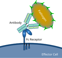
 示意图显示了Fc受体与抗体包被的微生物病原体的相互作用 | |
| 鑑定 | |
| 標誌 | Fc受体 |
| 膜蛋白數據庫 | 10 |
分类
编辑有几种不同类型的Fc受体,它们根据它们识别的抗体类型进行分类。与最常见的一类抗体IgG结合的称为Fcγ受体,与IgA结合的称为Fcα受体,与IgE结合的称为Fcε受体。Fc受体的类别还通过表达它们的细胞(巨噬细胞、粒细胞、自然杀伤细胞、T细胞和B细胞)和每个受体的信号传导特性来区分。[2]
Fcγ受体
编辑所有的Fcγ受体都属于免疫球蛋白超家族,是调理素(标记)微生物吞噬作用最重要的Fc受体。[3]该家族包括几个成员,Fcγ受体I(CD64)、Fcγ受体IIA(CD32)、Fcγ受体IIB(CD32)、Fcγ受体IIIA(CD16a)、Fcγ受体IIIB(CD16b),由于其不同的分子结构,它们的抗体亲和力不同。[4]例如,Fcγ受体I与IgG的结合比Fcγ受体II或Fcγ受体III更强。Fcγ受体I还具有由三个免疫球蛋白样结构域组成的细胞外部分,比Fcγ受体II或Fcγ受体III多一个结构域。这种特性允许Fcγ受体I结合单个IgG分子(或单体),但所有Fcγ受体必须结合免疫复合物中的多个IgG分子才能被激活。[5]
不同的Fcγ受体对IgG的亲和力各不相同,同样的,不同的IgG亚类对每种Fcγ受体都有独特的亲和力。这些相互作用由IgG CH2-84.4位置的聚糖(寡糖)进一步调节。 例如,通过产生位阻,含有CH2-84.4聚糖的岩藻糖会降低IgG对Fcγ受体IIIA的亲和力。相反,缺乏半乳糖并以GlcNAc部分终止的G0聚糖对Fcγ受体IIIA的亲和力会增加。
另一种Fc受体在多种细胞类型上表达,在结构上与MHC I类分子相似。该受体也结合IgG并参与该抗体的保存。[6]然而,由于这种Fc受体还参与将IgG从母亲通过胎盘转移到胎儿或通过乳汁转移到哺乳期婴儿,因此它被称为新生儿Fc受体(FcRn)。[7][8]最近,研究表明该受体在IgG血清水平的稳态中发挥作用。
Fcα受体
编辑只有一种Fc受体属于Fcα受体亚组,称为Fcα受体I(CD89)。[9]Fcα受体I存在于中性粒细胞、嗜酸性粒细胞、单核细胞、一些巨噬细胞(包括库普弗细胞)和一些树突状细胞的表面。[9]它由两个细胞外Ig样结构域组成,是免疫球蛋白超家族和多链免疫识别受体(MIRR)家族的成员。[3]它通过与两条Fc受体γ信号链结合发出信号。[9]另一种受体也可以结合IgA,尽管它对另一种称为IgM的抗体具有更高的亲和力。[10]这种受体称为Fcα/μ受体(Fcα/μR),是一种I型跨膜蛋白。这种Fc受体在其细胞外部分有一个Ig样结构域,它也是免疫球蛋白超家族的一员。[11]
Fcε受体
编辑已知有两种类型的Fcε受体:[3]
- 高亲和力受体Fcε受体I是免疫球蛋白超家族的成员(它有两个 Ig 样结构域)。Fcε受体I存在于表皮朗格汉斯细胞、嗜酸性粒细胞、肥大细胞和嗜碱性粒细胞中。[12][13]由于其细胞分布,该受体在控制过敏反应中起主要作用。Fcε受体I也在抗原呈递细胞上表达,并控制促炎性细胞因子的重要免疫介质的产生。[14]
- 低亲和力受体Fcε受体II(CD23)是一种C-型凝集素。Fcε受体II作为膜结合或可溶性受体具备多种功能:它能控制B细胞的生长和分化,并阻断嗜酸性粒细胞、单核细胞和嗜碱性粒细胞的IgE结合。[15]
汇总表
编辑| 受体名称 | 主要抗体配体 | 对配体的亲和力 | 细胞分布 | 与抗体结合后的效果 |
| Fcγ受体I(CD64) | IgG1和IgG3 | 高(Kd~10−9M) | 巨噬细胞中性粒细胞嗜酸性粒细胞树突状细胞 | 吞噬作用
细胞活化 激活呼吸爆发 诱导杀灭微生物 |
| Fcγ受体IIA(CD32) | IgG | 低(Kd>10−7M) | 巨噬细胞 | 吞噬作用
脱颗粒(嗜酸性粒细胞) |
| Fcγ受体IIB1(CD32) | IgG | 低(Kd>10−7M) | B细胞肥大细胞 | 无吞噬作用
抑制细胞活性 |
| Fcγ受体IIB2(CD32) | IgG | 低(Kd>10−7M) | 巨噬细胞
中性粒细胞 嗜酸性粒细胞 |
吞噬作用
抑制细胞活性 |
| Fcγ受体IIIA(CD16) | IgG | 低(Kd>10−6M) | 自然杀伤细胞巨噬细胞(某些组织) | 诱导抗体依赖的细胞介导的细胞毒性作用(ADCC)
巨噬细胞诱导细胞因子释放 |
| Fcγ受体IIIB(CD16) | IgG | 低(Kd>10−6M) | 嗜酸性粒细胞
巨噬细胞 中性粒细胞 肥大细胞 滤泡树突细胞 |
诱导杀灭微生物 |
| Fcε受体I | IgE | 高(Kd~10−10M) | 肥大细胞
嗜酸性粒细胞 嗜碱性粒细胞 朗格汉斯细胞 单核细胞 |
脱颗粒
吞噬作用 |
| Fcε受体II(CD23) | IgE | 低(Kd>10−7M) | B细胞
嗜酸性粒细胞 朗格汉斯细胞 |
有可能的粘附分子
IgE跨肠上皮细胞转运 增强过敏性致敏的正反馈机制(B细胞) |
| Fcα受体I(CD89) | IgA | 低(Kd>10−6M) | 单核细胞
巨噬细胞 中性粒细胞 嗜酸性粒细胞 |
吞噬作用
诱导杀灭微生物 |
| Fcα/μ受体(CD351) | IgA和IgM | IgM为高,IgA为中等 | B细胞
系膜细胞 巨噬细胞 |
胞吞作用诱导杀灭微生物 |
| Fcμ受体[16] | IgM | 未知 | 人类Fcμ受体主要由淋巴细胞表达,但不由吞噬细胞表达[17] | 功能尚未完全阐明/多样化[18] |
| 新生儿Fc受体 | IgG | 在酸性细胞内体,高
在pH中性细胞外环境,低 |
单核细胞 | 通过胎盘将IgG从母亲转移到胎儿
通过乳汁将IgG从母亲转移到婴儿 保护IgG免于降解 跨内皮/上皮层转移IgG |
作用
编辑Fc受体存在于免疫系统的许多细胞上,包括巨噬细胞和单核细胞等吞噬细胞、中性粒细胞和嗜酸性粒细胞等粒细胞,以及先天免疫系统(自然杀伤细胞)或适应性免疫系统(B细胞)的淋巴细胞。[19][20][21]它们允许这些细胞与附着在微生物或微生物感染细胞表面的抗体结合,并帮助这些细胞识别和消除微生物病原体。Fc受体在其Fc区(或尾部)结合抗体,这种相互作用可激活拥有Fc受体的细胞。[22]
激活吞噬细胞是Fc受体最常见的功能。例如,巨噬细胞在与Fcγ受体结合后开始通过吞噬作用摄取并杀死IgG包被的病原体。[23]另一个涉及Fc受体的过程称为抗体依赖的细胞介导的细胞毒性作用(ADCC)。在ADCC期间,自然杀伤细胞表面的Fcγ受体III刺激自然杀伤细胞从其颗粒体中释放细胞毒性分子以杀死抗体覆盖的细胞。[24]
Fcε受体I具有不一样的功能。Fcε受体I是粒细胞上的Fc受体,参与过敏反应和抵御寄生虫感染。当存在合适的过敏抗原或寄生虫时,至少两个IgE分子及其Fc受体在粒细胞表面的交联将触发细胞从其颗粒体中快速释放预先形成的介质。[3]
信号机制 - Fcγ受体
编辑激活
编辑Fcγ受体属于免疫受体,它们共享相似的信号通路(涉及酪氨酸残基磷酸化)。[25]这些受体通过称为免疫受体酪氨酸活化基序(ITAM)的重要激活基序在其细胞内产生信号。[26]ITAM是一种特定的氨基酸序列(YXXL),在受体的细胞内尾部连续出现两次。当磷酸盐基团通过Src家族激酶的膜锚定酶添加到ITAM的酪氨酸(Y)残基时,细胞内会产生信号级联。这种磷酸化反应通常发生在Fc受体与其配体相互作用之后。ITAM存在于Fcγ受体IIA的细胞内尾部,其磷酸化会诱导巨噬细胞的吞噬作用。Fcγ受体I和Fcγ受体IIIA没有ITAM,但可以通过与另一种具有ITAM的蛋白质相互作用将并激活信号传递给它们的吞噬细胞。这种衔接蛋白称为Fcγ亚基,与Fcγ受体IIA一样,包含ITAM特有的两个YXXL序列。
抑制
编辑仅存在一个YXXL基序不足以激活细胞,它代表一种基序(I/VXXYXXL),称为基于免疫受体酪氨酸的抑制基序(ITIM)。Fcγ受体IIB1和Fcγ受体IIB2具有ITIM序列,是抑制性Fc受体(它们不诱导吞噬作用)。这些受体的抑制作用受从酪氨酸残基上去除磷酸基团的酶控制,磷酸酶PTPN6和INPP5D抑制Fcγ受体的信号传导。[27]配体与Fcγ受体IIB的结合导致ITAM基序的酪氨酸磷酸化。这种修饰产生了磷酸酶的结合位点,一个SH2识别域。ITAM激活信号的取消是由Src家族蛋白酪氨酸激酶的抑制引起的,并且通过水解膜PIP3中断激活受体的进一步下游信号,例如激活Fcγ受体s、TCR、BCR 和细胞因子受体(例如c-Kit).[28]
Fcγ受体IIB的负信号传导对于活化B细胞的调节非常重要。阳性B细胞信号传导是由外来抗原与表面免疫球蛋白的结合启动的。分泌相同的抗原特异性抗体,它可以反馈抑制或促进负信号传导。这种负向信号由Fcγ受体IIB提供。[29]使用B细胞缺失突变体和显性失活酶的实验已经证明了含有SH2结构域的肌醇5-磷酸酶(SHIP)在负信号传导中的重要作用。通过SHIP的负信号似乎通过SH2结构域与Grb2和Shc的竞争抑制Ras通路,并且可能涉及细胞内脂质介质的消耗,这些介质充当变构酶激活剂或促进细胞外Ca2+的进入。[30]
细胞激活
编辑吞噬细胞
编辑当对某种抗原或表面成分具有特异性的IgG分子通过其Fab(抗原结合片段区)与病原体结合时,其Fc区指向外,可直接到达吞噬细胞。吞噬细胞将这些Fc区与其Fc受体结合。[23]许多低亲和力的相互作用在受体和抗体之间形成,它们共同作用以紧密结合抗体包被的微生物。低个体亲和力阻止Fc受体在没有抗原的情况下结合抗体,因此减少了在没有感染的情况下免疫细胞激活的机会。当没有抗原时,这也可以防止抗体对吞噬细胞的凝集。结合病原体后,抗体的Fc区与吞噬细胞的Fc受体之间的相互作用导致吞噬作用的启动。病原体通过涉及结合和释放Fc区或Fc受体复合物的活跃过程被吞噬细胞吞没,直到吞噬细胞的细胞膜完全包围病原体。[31]
自然杀伤细胞
编辑自然杀伤细胞上的Fc受体识别与病原体感染的靶细胞表面的IgG结合,称为CD16或Fcγ受体III。[32]IgG对Fcγ受体III的激活导致细胞因子(如II型干扰素)的释放,这些细胞因子向其他免疫细胞发出信号,而释放出的细胞毒性介质(如穿孔素和颗粒酶)会进入靶细胞并通过触发细胞凋亡促进细胞死亡。自然杀伤细胞上的Fcγ受体III也可以与单体IgG(即未与抗原结合的IgG)结合。发生这种情况时,Fc受体会抑制自然杀伤细胞的活性。[33]
肥大细胞
编辑IgE抗体与过敏原的抗原结合。这些过敏原结合的IgE分子与肥大细胞表面的Fcε受体进行相互作用。与Fcε受体I结合后,肥大细胞的激活会导致脱颗粒,肥大细胞由此从其细胞质颗粒中释放预制分子(包括组胺、蛋白聚糖和丝氨酸蛋白酶在内的化合物的混合物)。[34]活化的肥大细胞还会合成和分泌脂质衍生介质(如前列腺素、白三烯和血小板活化因子)和细胞因子(如白细胞介素-1、白细胞介素-3、白细胞介素-4、白细胞介素-5、白细胞介素-6、白细胞介素-13、肿瘤坏死因子、GM-CSF和几种趋化因子。)[35][36]这些介质通过吸引其他白细胞而导致炎症。
嗜酸性粒细胞
编辑曼氏血吸虫等大型寄生虫体型太大,无法被吞噬细胞吞噬。它们还有一个称为珠被的外部结构,可以抵抗巨噬细胞和肥大细胞释放的物质的攻击。然而,这些寄生虫可以被IgE包裹并被嗜酸性粒细胞表面的Fcε受体II识别。活化的嗜酸性粒细胞释放预先形成的介质,如主要碱性蛋白和酶(过氧化物酶),蠕虫对其不具有抗性。[37][38]Fcε受体II与蠕虫结合IgE的Fc部分的相互作用会导致嗜酸性粒细胞以类似于ADCC期间自然杀伤细胞的机制释放这些分子。[39]
T淋巴细胞
编辑CD4+ T细胞(辅助性T细胞)为产生抗体的B细胞提供帮助。在疾病病理学中观察到几个激活的效应CD4+ T细胞亚群。Sanders和Lynch在1993年总结的早期研究表明,Fc受体在CD4+ T细胞介导的免疫反应中起着关键作用,并提出在细胞表面的Fc受体和T细胞受体之间形成联合信号复合物。[40][41][42][43]Chauhan及其同事报告了标记的免疫复合物(IC)与活化的CD4+ T细胞表面上的CD3复合物共定位,因此表明Fc受体与T细胞受体复合物共存。[44]观察到这两种受体在活化的CD4+ T细胞膜上形成顶端结构,这表明这些受体的横向运动。[44]在细胞表面观察到Fc受体与T细胞受体和B细胞受体复合物的共迁移,T/B细胞偶联物在接触点显示这种共存。[45]较早的报告表明Fc受体在CD4+ T细胞上的表达是一个悬而未决的问题。[46]这确立了T细胞不表达Fc受体的当前范例,并且这些发现从未受到挑战和实验测试。[47]Chauhan及其同事展示IC与激活的CD4+ T细胞的Fc受体配体结合。[47]CD16a表达在活化的人类未成熟CD4+ T细胞中被诱导,这些细胞表达CD25、CD69和CD98,并且与IC的连接导致效应记忆细胞的产生。[48]CD16a信号由Syk(pSyk)的磷酸化介导。[48][49][50]
现在的一项研究表明,激活人类CD4+ T细胞后会诱导CD32a的表达,类似于CD16a。[51][52]HIV-1研究人员的三项独立研究也表明了CD4+ T细胞上的CD32a表达。CD16a和CD32a在活化的CD4+ T细胞亚群中的表达现已得到证实。[51][52]细胞表面的Fc受体在与由核酸组成的IC结合后触发细胞因子的产生并上调核酸传感途径。Fc受体存在于细胞表面和胞质溶胶中。CD16a信号上调核酸感应Toll样受体的表达,并将它们重新定位到细胞表面。[53][54]CD16a是人类CD4+ T细胞的一种新的共刺激信号,它成功地替代了自身免疫过程中的CD28需求。[55]在自身免疫背景下,CD4+ T细胞绕过CD28协同信号转导的要求而完全激活。[55]此外,CD28协同信号传导的阻断不会抑制TFH细胞的发育,TFH细胞是产生自身抗体的自身反应性浆B细胞的关键亚群。[56]免疫稳态需要共刺激信号和抑制信号之间的平衡。过度的共刺激或不充分的共抑制会导致耐受性崩溃和自身免疫。CD16a介导的共刺激在活化的CD4+ T细胞中提供阳性信号,而不是在缺乏Fcγ受体表达的静止细胞中。[51]
参见
编辑参考文献
编辑- ^ Anderson R. Manipulation of cell surface macromolecules by flaviviruses. Advances in Virus Research. 2003, 59: 229–74. ISBN 9780120398591. PMC 7252169 . PMID 14696331. doi:10.1016/S0065-3527(03)59007-8.
- ^ Owen J, Punt J, Stranford S, Jones P. Immunology 7th. New York: W.H. Freeman and Company. 2009: 423. ISBN 978-14641-3784-6.
- ^ 3.0 3.1 3.2 3.3 Fridman WH. Fc receptors and immunoglobulin binding factors. FASEB Journal. September 1991, 5 (12): 2684–90. PMID 1916092. S2CID 16805557. doi:10.1096/fasebj.5.12.1916092.
- ^ Indik ZK, Park JG, Hunter S, Schreiber AD. The molecular dissection of Fc gamma receptor mediated phagocytosis. Blood. December 1995, 86 (12): 4389–99. PMID 8541526. doi:10.1182/blood.V86.12.4389.bloodjournal86124389 .
- ^ Harrison PT, Davis W, Norman JC, Hockaday AR, Allen JM. Binding of monomeric immunoglobulin G triggers Fc gamma RI-mediated endocytosis. The Journal of Biological Chemistry. September 1994, 269 (39): 24396–402. PMID 7929100. doi:10.1016/S0021-9258(19)51097-3 .
- ^ Zhu X, Meng G, Dickinson BL, Li X, Mizoguchi E, Miao L, Wang Y, Robert C, Wu B, Smith PD, Lencer WI, Blumberg RS. MHC class I-related neonatal Fc receptor for IgG is functionally expressed in monocytes, intestinal macrophages, and dendritic cells. Journal of Immunology. March 2001, 166 (5): 3266–76. PMC 2827247 . PMID 11207281. doi:10.4049/jimmunol.166.5.3266.
- ^ Firan M, Bawdon R, Radu C, Ober RJ, Eaken D, Antohe F, Ghetie V, Ward ES. The MHC class I-related receptor, FcRn, plays an essential role in the maternofetal transfer of gamma-globulin in humans. International Immunology. August 2001, 13 (8): 993–1002. PMID 11470769. doi:10.1093/intimm/13.8.993 .
- ^ Simister NE, Jacobowitz Israel E, Ahouse JC, Story CM. New functions of the MHC class I-related Fc receptor, FcRn. Biochemical Society Transactions. May 1997, 25 (2): 481–6. PMID 9191140. doi:10.1042/bst0250481.
- ^ 9.0 9.1 9.2 Otten MA, van Egmond M. The Fc receptor for IgA (FcalphaRI, CD89). Immunology Letters. March 2004, 92 (1–2): 23–31. PMID 15081523. doi:10.1016/j.imlet.2003.11.018.
- ^ Shibuya A, Honda S. Molecular and functional characteristics of the Fcalpha/muR, a novel Fc receptor for IgM and IgA. Springer Seminars in Immunopathology. December 2006, 28 (4): 377–82. PMID 17061088. S2CID 23794895. doi:10.1007/s00281-006-0050-3.
- ^ Cho Y, Usui K, Honda S, Tahara-Hanaoka S, Shibuya K, Shibuya A. Molecular characteristics of IgA and IgM Fc binding to the Fcalpha/muR. Biochemical and Biophysical Research Communications. June 2006, 345 (1): 474–8 [2022-11-30]. PMID 16681999. doi:10.1016/j.bbrc.2006.04.084. hdl:2241/102010 . (原始内容存档于2018-11-03).
- ^ Ochiai K, Wang B, Rieger A, Kilgus O, Maurer D, Födinger D, Kinet JP, Stingl G, Tomioka H. A review on Fc epsilon RI on human epidermal Langerhans cells. International Archives of Allergy and Immunology. 1994,. 104 Suppl 1 (1): 63–4. PMID 8156009. doi:10.1159/000236756.
- ^ Prussin C, Metcalfe DD. 5. IgE, mast cells, basophils, and eosinophils. The Journal of Allergy and Clinical Immunology. February 2006, 117 (2 Suppl Mini–Primer): S450–6. PMID 16455345. doi:10.1016/j.jaci.2005.11.016.
- ^ von Bubnoff D, Novak N, Kraft S, Bieber T. The central role of FcepsilonRI in allergy. Clinical and Experimental Dermatology. March 2003, 28 (2): 184–7. PMID 12653710. S2CID 2080598. doi:10.1046/j.1365-2230.2003.01209.x.
- ^ Kikutani H, Yokota A, Uchibayashi N, Yukawa K, Tanaka T, Sugiyama K, Barsumian EL, Suemura M, Kishimoto T. Structure and function of Fc epsilon receptor II (Fc epsilon RII/CD23): a point of contact between the effector phase of allergy and B cell differentiation. Ciba Foundation Symposium. Novartis Foundation Symposia. 1989, 147: 23–31; discussion 31–5. ISBN 9780470513866. PMID 2695308. doi:10.1002/9780470513866.ch3.
- ^ FCMR Fc mu receptor [Homo sapiens (Human)] - Gene - NCBI. [2022-12-02]. (原始内容存档于2018-12-16).
- ^ Kubagawa H, Oka S, Kubagawa Y, Torii I, Takayama E, Kang DW, et al. Identity of the elusive IgMFc receptor (FcmuR) in humans. J. Exp. Med. 2009, 206 (12): 2779–93. PMC 2806608 . PMID 19858324. doi:10.1084/jem.20091107.
- ^ Liu J, Wang Y, Xiong E, Hong R, Lu Q, Ohno H, Wang JY. Role of the IgM Fc Receptor in Immunity and Tolerance. Frontiers in Immunology. 2019, 10: 529. PMC 6438924 . PMID 30967868. doi:10.3389/fimmu.2019.00529 .
- ^ Selvaraj P, Fifadara N, Nagarajan S, Cimino A, Wang G. Functional regulation of human neutrophil Fc gamma receptors. Immunologic Research. 2004, 29 (1–3): 219–30. PMID 15181284. S2CID 85351071. doi:10.1385/IR:29:1-3:219.
- ^ Sulica A, Chambers WH, Manciulea M, Metes D, Corey S, Rabinowich H, Whiteside TL, Herberman RB. Divergent signal transduction pathways and effects on natural killer cell functions induced by interaction of Fc receptors with physiologic ligands or antireceptor antibodies. Natural Immunity. 1995, 14 (3): 123–33. PMID 8832896.
- ^ Sarfati M, Fournier S, Wu CY, Delespesse G. Expression, regulation and function of human Fc epsilon RII (CD23) antigen. Immunologic Research. 1992, 11 (3–4): 260–72. PMID 1287120. S2CID 83698996. doi:10.1007/BF02919132.
- ^ Raghavan M, Bjorkman PJ. Fc receptors and their interactions with immunoglobulins (PDF). Annual Review of Cell and Developmental Biology. 1996, 12: 181–220. PMID 8970726. doi:10.1146/annurev.cellbio.12.1.181.
- ^ 23.0 23.1 Swanson JA, Hoppe AD. The coordination of signaling during Fc receptor-mediated phagocytosis. Journal of Leukocyte Biology. December 2004, 76 (6): 1093–103. PMID 15466916. S2CID 13912335. doi:10.1189/jlb.0804439. hdl:2027.42/141562 .
- ^ Sun PD. Structure and function of natural-killer-cell receptors. Immunologic Research. 2003, 27 (2–3): 539–48 [2022-12-03]. PMID 12857997. S2CID 29921323. doi:10.1385/IR:27:2-3:539. (原始内容存档于2022-12-03).
- ^ Dushek O, Goyette J, van der Merwe PA. Non-catalytic tyrosine- phosphorylated receptors. Immunological Reviews. November 2012, 250 (1): 258–276. PMID 23046135. S2CID 1549902. doi:10.1111/imr.12008.
- ^ Cambier JC. New nomenclature for the Reth motif (or ARH1/TAM/ARAM/YXXL). Immunology Today. February 1995, 16 (2): 110. PMID 7888063. doi:10.1016/0167-5699(95)80105-7.
- ^ Huang ZY, Hunter S, Kim MK, Indik ZK, Schreiber AD. The effect of phosphatases SHP-1 and SHIP-1 on signaling by the ITIM- and ITAM-containing Fcgamma receptors FcgammaRIIB and FcgammaRIIA. Journal of Leukocyte Biology. June 2003, 73 (6): 823–9. PMID 12773515. S2CID 14502303. doi:10.1189/jlb.0902454 .
- ^ Cambier JC. Inhibitory receptors abound?. Proceedings of the National Academy of Sciences of the United States of America. June 1997, 94 (12): 5993–5. Bibcode:1997PNAS...94.5993C. PMC 33673 . PMID 9177155. doi:10.1073/pnas.94.12.5993 .
- ^ Takai T, Ono M, Hikida M, Ohmori H, Ravetch JV. Augmented humoral and anaphylactic responses in Fc gamma RII-deficient mice. Nature. January 1996, 379 (6563): 346–9. Bibcode:1996Natur.379..346T. PMID 8552190. S2CID 4364705. doi:10.1038/379346a0.
- ^ Coggeshall KM. Inhibitory signaling by B cell Fc gamma RIIb. Current Opinion in Immunology. June 1998, 10 (3): 306–12. PMID 9638367. doi:10.1016/s0952-7915(98)80169-6.
- ^ Joshi T, Butchar JP, Tridandapani S. Fcgamma receptor signaling in phagocytes. International Journal of Hematology. October 2006, 84 (3): 210–6. PMID 17050193. S2CID 6501210. doi:10.1532/IJH97.06140.
- ^ Trinchieri G, Valiante N. Receptors for the Fc fragment of IgG on natural killer cells. Natural Immunity. 1993, 12 (4–5): 218–34. PMID 8257828.
- ^ Sulica A, Galatiuc C, Manciulea M, Bancu AC, DeLeo A, Whiteside TL, Herberman RB. Regulation of human natural cytotoxicity by IgG. IV. Association between binding of monomeric IgG to the Fc receptors on large granular lymphocytes and inhibition of natural killer (NK) cell activity. Cellular Immunology. April 1993, 147 (2): 397–410. PMID 8453679. doi:10.1006/cimm.1993.1079.
- ^ Yamasaki S, Saito T. Regulation of mast cell activation through FcepsilonRI. Chemical Immunology and Allergy. 2005, 87: 22–31. ISBN 3-8055-7948-9. PMID 16107760. doi:10.1159/000087568.
- ^ Wakahara S, Fujii Y, Nakao T, Tsuritani K, Hara T, Saito H, Ra C. Gene expression profiles for Fc epsilon RI, cytokines and chemokines upon Fc epsilon RI activation in human cultured mast cells derived from peripheral blood. Cytokine. November 2001, 16 (4): 143–52. PMID 11792124. doi:10.1006/cyto.2001.0958.
- ^ Metcalfe DD, Baram D, Mekori YA. Mast cells. Physiological Reviews. October 1997, 77 (4): 1033–79. PMID 9354811. doi:10.1152/physrev.1997.77.4.1033.
- ^ David JR, Butterworth AE, Vadas MA. Mechanism of the interaction mediating killing of Schistosoma mansoni by human eosinophils. The American Journal of Tropical Medicine and Hygiene. September 1980, 29 (5): 842–8. PMID 7435788. doi:10.4269/ajtmh.1980.29.842.
- ^ Capron M, Soussi Gounni A, Morita M, Truong MJ, Prin L, Kinet JP, Capron A. Eosinophils: from low- to high-affinity immunoglobulin E receptors. Allergy. 1995, 50 (25 Suppl): 20–3. PMID 7677229. S2CID 36197719. doi:10.1111/j.1398-9995.1995.tb04270.x.
- ^ Gounni AS, Lamkhioued B, Ochiai K, Tanaka Y, Delaporte E, Capron A, Kinet JP, Capron M. High-affinity IgE receptor on eosinophils is involved in defence against parasites. Nature. January 1994, 367 (6459): 183–6. Bibcode:1994Natur.367..183S. PMID 8114916. S2CID 4331405. doi:10.1038/367183a0.
- ^ Pichler WJ, Lum L, Broder S. Fc-receptors on human T lymphocytes. I. Transition of Tgamma to Tmu cells. J Immunol. 1978, 121 (4): 1540–1548. PMID 308968.
- ^ Sandor M, Lynch RG. Lymphocyte Fc receptors: the special case of T cells. Immunol. Today. May 1993, 14 (5): 227–31. PMID 8517922. doi:10.1016/0167-5699(93)90168-K.
- ^ Engelhardt W, Matzke J, Schmidt RE. Activation-dependent expression of low affinity IgG receptors Fc gamma RII(CD32) and Fc gamma RIII(CD16) in subpopulations of human T lymphocytes. Immunobiology. 1995, 192 (5): 297–320. PMID 7649565. doi:10.1016/s0171-2985(11)80172-5.
- ^ Moretta L, Webb SR, Grossi CE, Lydyard PM, Cooper MD. Functional analysis of two human T-cell subpopulations: help and suppression of B-cell responses by T cells bearing receptors for IgM or IgG. J Exp Med. 1977, 146 (1): 184–200. PMC 2180738 . PMID 301544. doi:10.1084/jem.146.1.184.
- ^ 44.0 44.1 Chauhan AK, Moore TL. T cell activation by terminal complex of complement and immune complexes. The Journal of Biological Chemistry. 2011, 286 (44): 38627–38637. PMC 3207419 . PMID 21900254. doi:10.1074/jbc.M111.266809 .
- ^ Sandor M, Lynch RG. Lymphocyte Fc receptors: the special case of T cells. Immunology Today. 1993, 14 (5): 227–231. PMID 8517922. doi:10.1016/0167-5699(93)90168-K.
- ^ Nimmerjahn F, Ravetch JV. Fcgamma receptors as regulators of immune responses. Nat. Rev. Immunol. January 2008, 8 (1): 34–47. PMID 18064051. S2CID 34597359. doi:10.1038/nri2206.
- ^ 47.0 47.1 Bruhns P, Jönsson F. Mouse and human FcR effector functions. Immunol. Rev. November 2015, 268 (1): 25–51. PMID 26497511. S2CID 19544801. doi:10.1111/imr.12350.
- ^ 48.0 48.1 Chauhan AK, Moore TL, Bi Y, Chen C. FcγRIIIa-Syk Co-signal Modulates CD4+ T-cell Response and Up-regulates Toll-like Receptor (TLR) Expression. J. Biol. Chem. January 2016, 291 (3): 1368–86. PMC 4714221 . PMID 26582197. doi:10.1074/jbc.M115.684795 .
- ^ Chauhan AK, Chen C, Moore TL, DiPaolo RJ. Induced expression of FcγRIIIa (CD16a) on CD4+ T cells triggers generation of IFN-γhigh subset. J. Biol. Chem. February 2015, 290 (8): 5127–40. PMC 4335247 . PMID 25556651. doi:10.1074/jbc.M114.599266 .
- ^ Chauhan AK, Moore TL. Immune complexes and late complement proteins trigger activation of Syk tyrosine kinase in human CD4(+) T cells. Clin Exp Immunol. 2012, 167 (2): 235–245. PMC 3278689 . PMID 22235999. doi:10.1111/j.1365-2249.2011.04505.x.
- ^ 51.0 51.1 51.2 Chauhan AK, Chen C, Moore TL, DiPaolo RJ. Induced expression of FcγRIIIa (CD16a) on CD4+ T cells triggers generation of IFN-γhigh subset. J. Biol. Chem. February 2015, 290 (8): 5127–40. PMC 4335247 . PMID 25556651. doi:10.1074/jbc.M114.599266 .
- ^ 52.0 52.1 Holgado MP, Sananez I, Raiden S, Geffner JR, Arruvito L. CD32 Ligation Promotes the Activation of CD4+ T Cells. Front Immunol. 2018, 9: 2814. PMC 6284025 . PMID 30555482. doi:10.3389/fimmu.2018.02814 .
- ^ Chauhan AK, Moore TL, Bi Y, Chen C. FcγRIIIa-Syk Co-signal Modulates CD4+ T-cell Response and Up-regulates Toll-like Receptor (TLR) Expression. J. Biol. Chem. January 2016, 291 (3): 1368–86. PMC 4714221 . PMID 26582197. doi:10.1074/jbc.M115.684795 .
- ^ Chauhan AK. FcgammaRIIIa Signaling Modulates Endosomal TLR Responses in Human CD4+ T Cells. J Immunol. 2017, 198 (12): 4596–4606. PMC 5505339 . PMID 28500073. doi:10.4049/jimmunol.1601954.
- ^ 55.0 55.1 Bour-Jordan H, Esensten JH, Martinez-Llordella M, Penaranda C, Stumpf M, Bluestone JA. Intrinsic and extrinsic control of peripheral T-cell tolerance by costimulatory molecules of the CD28/ B7 family. Immunol Rev. 2011, 241 (1): 180–205. PMC 3077803 . PMID 21488898. doi:10.1111/j.1600-065X.2011.01011.x.
- ^ Weber JP, Fuhrmann F, Feist RK, Lahmann A, Al Baz MS, Gentz LJ, Vu Van D, Mages HW, Haftmann C, Riedel R, Grun JR, Schuh W, Kroczek RA, Radbruch A, Mashreghi MF, Hutloff A. ICOS maintains the T follicular helper cell phenotype by down-regulating Kruppel-like factor 2. The Journal of Experimental Medicine. 2015, 212 (2): 217–233. PMC 4322049 . PMID 25646266. doi:10.1084/jem.20141432.
拓展阅读
编辑- Janeway CA, Travers P, Waldport M, Shlomchik MJ. Chapter 9. The Humoral Immune Response. Immunobiology: The Immune System in Health and Disease 5th. New York: Garland. 2001. ISBN 978-0-8153-3642-6.
- Abbas AK, Lichtman AH, Pillai S. Chapter 12: Effector Mechanisms of Humoral Immunity. Cellular and molecular immunology 7th. Philadelphia: Elsevier/Saunders. 2012. ISBN 978-1-4377-1528-6.
- Gerber JS, Mosser DM. Stimulatory and inhibitory signals originating from the macrophage Fcgamma receptors. Microbes and Infection. February 2001, 3 (2): 131–9. PMID 11251299. doi:10.1016/s1286-4579(00)01360-5.
- Maverakis E, Kim K, Shimoda M, Gershwin ME, Patel F, Wilken R, Raychaudhuri S, Ruhaak LR, Lebrilla CB. Glycans in the immune system and The Altered Glycan Theory of Autoimmunity: a critical review. Journal of Autoimmunity. February 2015, 57: 1–13. PMC 4340844 . PMID 25578468. doi:10.1016/j.jaut.2014.12.002.